IMAGE

Fig. 2

ID
ZDB-IMAGE-200812-2
Source
Figures for Shen et al., 2020
Image
Figure Caption

Fig. 2

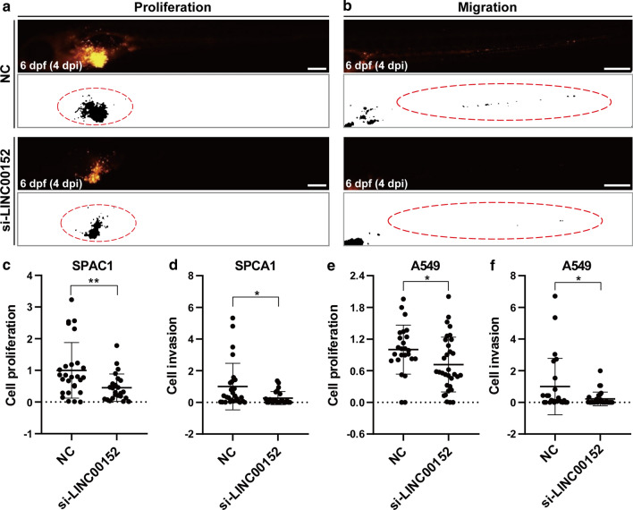
Knockdown of LINC00152 decreases the proliferation and invasion of lung cancer cells in zebrafish xenograft by stereomicroscopy. a, b Lung cancer cells transfected with si-LINC00152 siRNA or NC were injected into the PVS of 2-dpf WT zebrafish larvae. Images were taken using a stereomicroscope at 4 dpi. CM-DiI-positive areas in the yolk were quantified for proliferation (a), and CM-DiI-positive areas in trunk were quantified for invasion (b). The regions enclosed by the red dashed curve in the segmented images were selected for calculating tumor areas in the yolk or trunk. c, d Statistical analysis of proliferation (c) and invasion (d) when knocking down LINC00152 in SPCA1 cells. e, f Statistical analysis of proliferation (c) and invasion (d) when knocking down LINC00152 in A549 cells. *: p < 0.05, **: p < 0.01. Scale: 250 μm

Acknowledgments
This image is the copyrighted work of the attributed author or publisher, and ZFIN has permission only to display this image to its users. Additional permissions should be obtained from the applicable author or publisher of the image. Full text @ Cancer Cell Int.