
- Title
-
Preclinical evaluation of platinum-loaded hydroxyapatite nanoparticles in an embryonic zebrafish xenograft model
- Authors
- Nadar, R.A., Asokan, N., Degli Esposti, L., Curci, A., Barbanente, A., Schlatt, L., Karst, U., Iafisco, M., Margiotta, N., Brand, M., van den Beucken, J.J.J.P., Bornhäuser, M., Leeuwenburgh, S.C.G.
- Source
- Full text @ Nanoscale

Effect of direct addition or co-injection of PtPP on zebrafish embryo hosting breast cancer cells. (A) Representative images of zebrafish embryo expressing vascular marker Tg(kdrl:Has.HRAS-mCherry) in casper background hosting eGFP labeled breast cancer cells (MDA-MB-231_eGFP) at 2 days post injection: in contrast to untreated Pt-free controls, direct addition of 30 μM PtPP (center) and co-injection of 20 μM PtPP (bottom) revealed a reduction in breast cancer cell number. Vasculature is indicated in magenta and breast cancer cells are depicted in green. White arrowheads correspond to presence of cancer cells in the respective regions. Scale bar: 500 μm. (B) Manual quantification of cancer cells at 2 days post injection revealing a significant reduction of breast cancer cell number in PtPP-treated embryos compared to controls. Plot represents mean ± SEM. Statistical analysis: one-way ANOVA followed by Dunnett's test for multiple comparisons. ****P < 0.0001. |

Biodistribution of fluorescently labeled HA-Cit and HA nanoparticles in zebrafish embryo. Biodistribution of fluorescently labeled HA-nanoparticles (in red) injected in casper embryos at 2 days post injection (2 dpi). At concentrations between 150–500 μg ml−1, all citrate-functionalized HA-Cit nanoparticles (top) were spread homogeneously throughout the embryos, whereas citrate-free HA nanoparticles accumulated near the injection site (bottom). HA nanoparticles are depicted in red, white arrowheads correspond to fluorescently labeled HA nanoparticles. Scale bar: 500 μm. |
Effect of PtPP-loaded and citrate-functionalized HA nanoparticles (PtPP-HA-Cit) nanoparticles on survival of breast cancer cells in vivo. (A) Zebrafish embryo expressing vascular marker Tg(kdrl:Has.HRAS-mCherry) in casper background injected with eGFP-labeled breast cancer cells (MDA-MB-231_eGFP) at 2 days post injection. Co-injection of 300 μg ml−1 of PtPP-loaded HA nanoparticles and breast cancer cells (bottom) decreased cancer cell numbers as compared to untreated controls (top). Vasculature is shown in magenta and breast cancer cells are depicted in green. White arrowheads indicate the presence of cancer cells. Scale bar: 500 μm. (B) Manual quantification of cancer cell number at 2 days post injection indicating that PtPP-loaded HA nanoparticles reduced survival of breast cancer cells in zebrafish embryos as compared to untreated controls. Plot represents mean ± SEM. Statistical analysis: two-tailed Mann Whitney U Test. **P < 0.01. |

Breast cancer cells in xenografted zebrafish embryos. A) Representative image of zebrafish embryos expressing vascular marker Tg(kdrl:Has.HRAS-mCherry) in casper background hosting eGFP labeled breast cancer cells (MDA-MB-231_eGFP) at 24 h post injection. Cancer cells injected at the Duct of Cuvier (blue arrowhead) migrated throughout the embryo (white arrowhead). Vasculature is indicated in magenta and breast cancer cells are depicted in green. Scale bar: 500 μm. B) Representative image of breast cancer cells in the caudal tail region (left, white arrowhead) of the embryo exhibiting extravasation by forming protrusion (right, white arrowhead). Scale bar: 50 μm. |
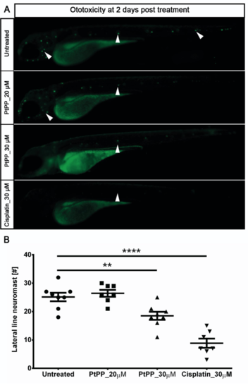
Ototoxicity assay in zebrafish embryos. A) Embryos were treated with PtPP (20 μM and 30 μM) and Cisplatin (positive control, 30 μM) and lateral line neuromasts were stained using DASPEI. Live imaging of these treated, stained embryos showed ototoxic effect of PtPP on the neuromast cells as expressed by a reduced amount of these cells caused by 30 μM of PtPP and cisplatin treatments after 2 days post treatment (neuromast cells indicated with white arrowheads). B) Quantification of the lateral line neuromast cells stained by DASPEI after 2 days post treatment with PtPP and cisplatin. Treatment using 30 μM cisplatin and 30 μM PtPP caused a significant reduction of neuromast cells. The plot represents means ± sem. **0.001 <P < 0.01; ****P <0.0001. |
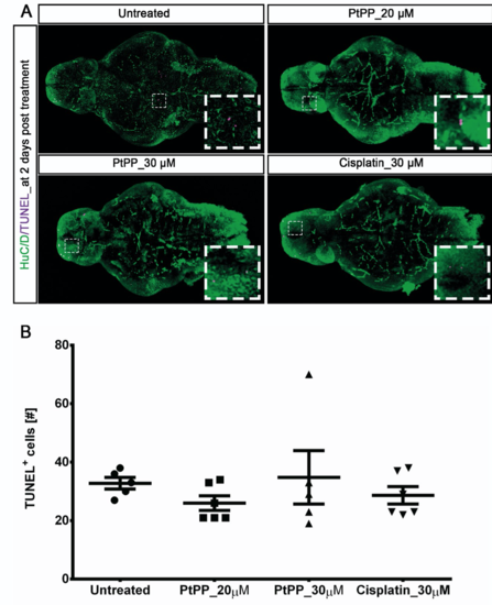
Cell death in zebrafish larvae brains due to PtPP treatment. A) After PtPP and cisplatin treatments, larvae brains were cleared and stained for apoptotic cells using the TUNEL assay. Confocal imaging of these stained larvae brains showed a basal level of apoptotic cells irrespective of PtPP and cisplatin treatments (indicated by white dashed line boxes pointing at TUNEL+ cells in the brain). Neurons are stained with HuC/D (green) and apoptotic cells are stained with TUNEL (magenta). Inserts show images at higher magnifications of the areas indicated with a white dashed line box. B) Quantification of the TUNEL+ cells in the zebrafish larvae brain after 2 days post treatment with PtPP and cisplatin. Cisplatin and PtPP treatments did not show any neurotoxic effect. |