
|
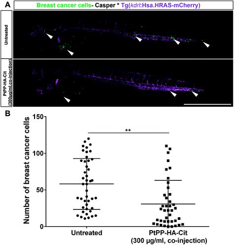
Fig. 5
Effect of PtPP-loaded and citrate-functionalized HA nanoparticles (PtPP-HA-Cit) nanoparticles on survival of breast cancer cells in vivo. (A) Zebrafish embryo expressing vascular marker Tg(kdrl:Has.HRAS-mCherry) in casper background injected with eGFP-labeled breast cancer cells (MDA-MB-231_eGFP) at 2 days post injection. Co-injection of 300 μg ml−1 of PtPP-loaded HA nanoparticles and breast cancer cells (bottom) decreased cancer cell numbers as compared to untreated controls (top). Vasculature is shown in magenta and breast cancer cells are depicted in green. White arrowheads indicate the presence of cancer cells. Scale bar: 500 μm. (B) Manual quantification of cancer cell number at 2 days post injection indicating that PtPP-loaded HA nanoparticles reduced survival of breast cancer cells in zebrafish embryos as compared to untreated controls. Plot represents mean ± SEM. Statistical analysis: two-tailed Mann Whitney U Test. **P < 0.01.