FIGURE SUMMARY
Title

Elongator Subunit 3 (Elp3) Is Required for Zebrafish Trunk Development

Authors
Rojas-Benítez, D., L Allende, M.
Source
Full text @ Int. J. Mol. Sci.

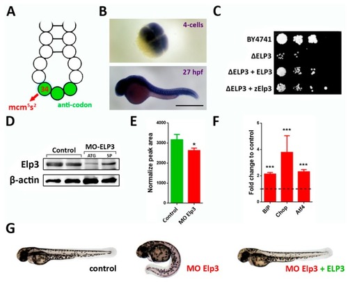
Elp3 morphants present a ventrally-curved tail. (A) Schematic representation of the anti-codon loop of a tRNA, indicating position 34 (red arrow) where 5-methoxycarbonylmethyl-2-thiouridine (mcm5s2U) is located. Anti-codon is depicted in green. (B) In situ hybridization of Elp3 mRNA in 4 cell (upper) and 27 hpf (lower) embryos. Scale bar 0.7 mm. (C) Saccharomyces cerevisiae mutants for ELP3 (ΔELP3) were transformed with an expression plasmid that contained zebrafish Elp3 (zElp3). BY4741 was used as control. Image representative of three independent experiments. (D) Western blot detecting Elp3 in 48 hpf samples from control and morphant embryos (MO-Elp3); β-actin was used as loading control. Samples from embryos injected with translation- (ATG) and splicing-blocking (SP). (E) Liquid chromatography-mass spectrometry (LC-MS) analysis of tRNA modification. Peak area values for mcm5s2U are shown normalized to the of purified tRNA for each sample (n = 3, * p < 0.05). (F) qPCR for UPR targets BiP, Chop and Atf4. Fold change in mRNA levels comparing morphant embryos to controls; t-test was performed for statistical analysis. *** p < 0.005). (G) Representative images of control, morphants and MO + ELP3 mRNA injected embryos by 48 hpf (images representative of at least six independent experiments, in which at least 100 embryos were analyzed).

EXPRESSION / LABELING:
Gene:
Fish:
Knockdown Reagents:
Anatomical Term:
Stage Range: 4-cell to Long-pec
PHENOTYPE:
Fish:
Knockdown Reagents:
Observed In:
Stage: Long-pec

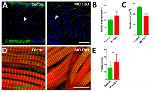
Aberrant somite shape and horizontal myoseptum in Elp3 morphants. To show somite boundary we detected β-dystroglycan in (A) control and morphant 48 hpf embryos, nuclei were stained with TO-PRO. Images representative of three independent experiments. Horizontal myoseptum is indicated with a white arrowhead. Scale Bar 50 µm. Also, somite angle (B) and area (C) were measured (N = 3, n = 50, t-Student. *** p < 0.005). Myomesin was also detected (D) in 48 hpf control and morphant embryos (N = 3, n = 50, t-Student). Scale bar 10 µm. (E) To evaluate muscle function coiling was measured counting twitches per minute in 30 hpf embryos (N = 3, n = 50, t-Student; ns non-significant).

Abnormal muscle fiber morphology in morphants. Muscle fibers of control (A, D, G and J), morphants (B, E, H and K) and MO + PKADN mRNA injected (C, F, I and L) were analyzed. Upper panels show fast fibers (F59) and lower show slow fibers (S58). F-actin was stained with TRICT-phalloidin. Scale bar 50 µm. Images representative of 4 independent experiments.

EXPRESSION / LABELING:
Antibodies:
Fish:
Knockdown Reagents:
Anatomical Terms:
Stage: Long-pec

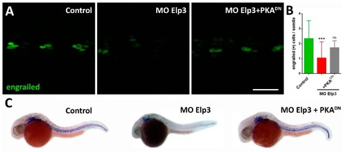
Shh activity is diminished in ELP morphants. (A) Immunohistochemistry usnig an anti-Engrailed antibody to visualize the adaxial cells. Control, elp3 morphants and MO/PKADN mRNA-injected animals were analyzed 24 hpf. (B) The number of engrailed(+) cells were counted (N = 4, n = 30, ANOVA *** p < 0.005). (C) In situ hybridization to detect the expression pattern of col2a1 in control, elp3 morphants and MO/PKADN mRNA-injected fish (images representative of four independent experiments). Scale bar 40 µm.

EXPRESSION / LABELING:
Gene:
Antibody:
Fish:
Knockdown Reagents:
Anatomical Terms:
Stage: Prim-5
PHENOTYPE:
Fish:
Knockdown Reagents:
Observed In:
Stage: Prim-5

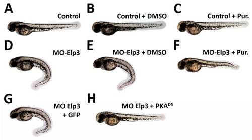
Morphant phenotype is rescued by sonic hedgehog pathway activation. Images of 2dpf larvae of (A) Control, (B) Control + 0.1% DMSO (C) Control + Pur 10 µM, (D) MO-Elp3 injected, (E) MO-Elp3 + 0.1% DMSO and (F) MO-Elp3 + Pur 10 µM. (G) MO Elp3 + GFP mRNA and (H) MO Elp3 + PKADN mRNA. Images are representative of four independent experiments.

Acknowledgments
This image is the copyrighted work of the attributed author or publisher, and ZFIN has permission only to display this image to its users. Additional permissions should be obtained from the applicable author or publisher of the image. Full text @ Int. J. Mol. Sci.