IMAGE

Figure 2

ID
ZDB-IMAGE-200314-57
Source
Figures for Rojas-Benítez et al., 2020
Image
Figure Caption

Figure 2

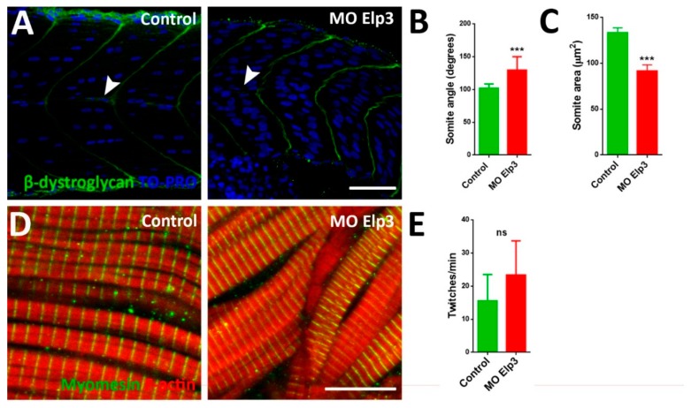
Aberrant somite shape and horizontal myoseptum in Elp3 morphants. To show somite boundary we detected β-dystroglycan in (A) control and morphant 48 hpf embryos, nuclei were stained with TO-PRO. Images representative of three independent experiments. Horizontal myoseptum is indicated with a white arrowhead. Scale Bar 50 µm. Also, somite angle (B) and area (C) were measured (N = 3, n = 50, t-Student. *** p < 0.005). Myomesin was also detected (D) in 48 hpf control and morphant embryos (N = 3, n = 50, t-Student). Scale bar 10 µm. (E) To evaluate muscle function coiling was measured counting twitches per minute in 30 hpf embryos (N = 3, n = 50, t-Student; ns non-significant).

Figure Data
Acknowledgments
This image is the copyrighted work of the attributed author or publisher, and ZFIN has permission only to display this image to its users. Additional permissions should be obtained from the applicable author or publisher of the image. Full text @ Int. J. Mol. Sci.