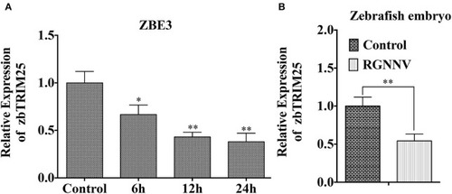
zbTRIM25 expression is inhibited in zebrafish during RGNNV infection. (A) ZBE3 cells were infected with RGNNV for the indicated time points. (B) Zebrafish embryos were infected with RGNNV for 24 h. The expression of zbTRIM25 mRNA was tested by qRT-PCR and normalized with 18s rRNA. Asterisks indicate significant differences between groups (*p < 0.05; **p < 0.01).

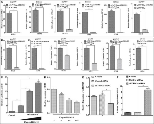
zbTRIM25 potentiates RLR signaling pathway. (A,B) ZBE3 cells were transfected with pCMV-Flag-zbTRIM25 or pCMV-Flag vector for 24 h. After treated with RGNNV (A) or poly I:C (B), the transcript levels of RIG-I, MAVS, TRAF3, IRF3, IFN 1, and ISG15 mRNA in ZBE3 cells were analyzed. (C) HEK 293T cells were transfected with pCMV-Myc or pCMV-Myc-zbRIG-I together with pCMV-Flag or increasing amount of pCMV-Flag-zbTRIM25 as well as DrIFN1 pro-Luc and pRL-TK. Luciferase activities were measured and normalized to the amount of Renilla Luciferase activities. (D) ZBE3 cells were transfected with pCMV-Flag and increasing amount of pCMV-Flag-zbTRIM25 for 24 h, and then infected with RGNNV for 24 h. QRT-PCR analysis was performed for RDRP. (E,F) ZBE3 cells were either not transfected (Control) or transfected with 100 nM Control siRNA (NC) or 100 nM zbTRIM25 siRNA. Cells were then subcultured for 24 h and infected with RGNNV for 24 h. Cells were harvested subsequently. Levels of zbTRIM25 and RDRP mRNA were analyzed by qRT-PCR and normalized with 18s rRNA. Data represent the mean + SD (n = 3). Asterisks indicate significant differences between groups (*p < 0.05; **p < 0.01).

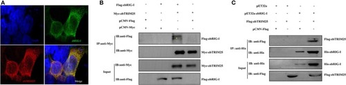
zbTRIM25 interacts with zbRIG-I. (A) HEK 293T cells were transfected with pCMV-Flag-zbRIG-I and pCMV-Myc-zbTRIM25 plasmids for 24 h, and then cells were immunostained with anti-Myc and anti-Flag antibodies and analyzed by fluorescence microscopy. All nuclei were stained with Hoechst 33342. (B) HEK 293T cells were transfected with plasmids as indicated for 24 h. After treated with poly I:C for 24 h, cells were lysed, and the cell lysates were either analyzed directly by using anti-Myc and anti-Flag antibodies via Western blotting (Input) or subjected to immunoprecipitation using anti-Myc antibodies. The precipitates (IP) were analyzed by Western blotting with anti-Myc and anti-Flag antibodies, respectively. (C) The purified His-zbRIG-I proteins were incubated with the lysates of HEK 293T cells transfected with the indicated plasmids, respectively. The pulled down proteins and original cell lysates were examined by Western blotting with indicated antibodies.

Physical interaction of zbTRIM25 with zbRIG-I. (A) Schematic representation of full-length zbRIG-I and zbRIG-I deletion mutants. (B–E) Interactions of GFP-tagged zbRIG-I-2CARD (B), zbRIG-I-Δ2CARD (C), zbRIG-I-RD (D), or zbRIG-I-Δ(CARDs+RD) (E) with Flag-tagged zbTRIM25 were examined using immunoprecipitation assays. HEK 293T cells were transfected with plasmids as indicated for 24 h. After treated with poly I:C for 24 h, cells were lysed, and the cell lysates were either analyzed directly by using anti-GFP and anti-Flag antibodies via Western blotting (Input) or subjected to immunoprecipitation using anti-GFP antibodies. The precipitates (IP) were analyzed by Western blotting with anti-GFP and anti-Flag antibodies, respectively. (F) Schematic illustration of zbTRIM25 truncations. Interactions of GFP-zbRIG-I-RD with Flag-zbTRIM25-ΔSPRY (G) or Flag-zbTRIM25-SPRY (H), GFP-zbRIG-I-2CARD with Flag-zbTRIM25-ΔSPRY (I), or Flag-zbTRIM25-SPRY (J) were examined using immunoprecipitation assays. HEK 293T cells were transfected with plasmids as indicated for 24 h. Cells were lysed after treated with poly I:C for 24 h. Immunoprecipitation and immunoblotting were performed with indicated antibodies.

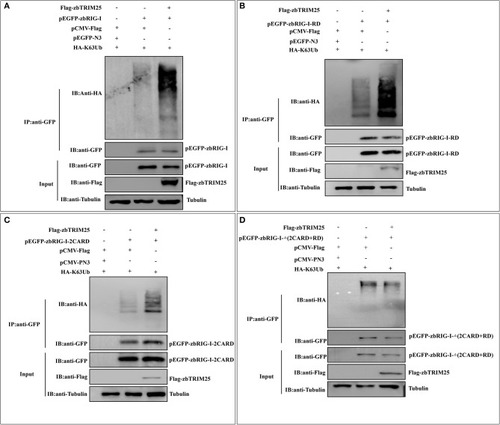
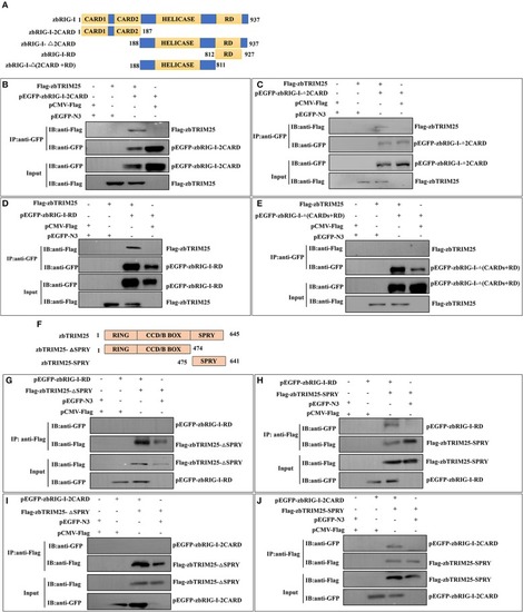
zbTRIM25 promotes zbRIG-I ubiquitination. (A–D) HEK 293T cells were transfected with plasmids as indicated for 24 h. At 24 h after poly I:C treatment, cells were lysed, and the cell lysates were either analyzed directly by using anti-GFP, anti-Flag, and anti-tubulin antibodies via Western blotting (Input) or subjected to immunoprecipitation using anti-GFP antibodies. The precipitates (IP) were analyzed by Western blotting with anti-GFP and anti-HA antibodies, respectively.

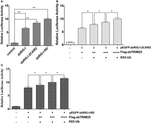
zbTRIM25-mediated K63 ubiquitination in 2CARD and RD regions of zbRIG-I is important for IFN induction. (A) HEK 293T cells were transfected with pEGFP-zbRIG-I, pEGFP-zbRIG-I-2CARD, and pEGFP-zbRIG-I-RD as well as DrIFN1 pro-Luc and pRL-TK vectors. (B,C) HEK 293T cells were transfected with pEGFP-zbRIG-I-2CARD or pEGFP-zbRIG-I-RD together with HA-K63Ub and increasing amount of pCMV-Flag-zbTRIM25 as well as DrIFN1 pro-Luc and pRL-TK plasmids. Luciferase activities were measured and normalized to the amount of Renilla Luciferase activities. Data represent the mean + SD (n = 3). Asterisks indicate significant differences between groups (*p < 0.05; **p < 0.01).

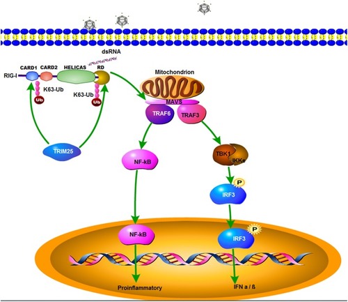
A schematic model of zbTRIM25-mediated RLR signaling pathway during RGNNV infection. zbTRIM25 interacts with and catalyzes the K63 polyubiquitination of 2CARD and RD regions of zbRIG-I, which subsequently induces the activation of downstream signaling event via MAVS, and thereby inhibits viral infection.

Acknowledgments
This image is the copyrighted work of the attributed author or publisher, and ZFIN has permission only to display this image to its users. Additional permissions should be obtained from the applicable author or publisher of the image. Full text @ Front Immunol