IMAGE

Figure 3

ID
ZDB-IMAGE-191230-1238
Source
Figures for Jin et al., 2019
Image
Figure Caption

Figure 3

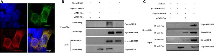
zbTRIM25 interacts with zbRIG-I. (A) HEK 293T cells were transfected with pCMV-Flag-zbRIG-I and pCMV-Myc-zbTRIM25 plasmids for 24 h, and then cells were immunostained with anti-Myc and anti-Flag antibodies and analyzed by fluorescence microscopy. All nuclei were stained with Hoechst 33342. (B) HEK 293T cells were transfected with plasmids as indicated for 24 h. After treated with poly I:C for 24 h, cells were lysed, and the cell lysates were either analyzed directly by using anti-Myc and anti-Flag antibodies via Western blotting (Input) or subjected to immunoprecipitation using anti-Myc antibodies. The precipitates (IP) were analyzed by Western blotting with anti-Myc and anti-Flag antibodies, respectively. (C) The purified His-zbRIG-I proteins were incubated with the lysates of HEK 293T cells transfected with the indicated plasmids, respectively. The pulled down proteins and original cell lysates were examined by Western blotting with indicated antibodies.

Acknowledgments
This image is the copyrighted work of the attributed author or publisher, and ZFIN has permission only to display this image to its users. Additional permissions should be obtained from the applicable author or publisher of the image. Full text @ Front Immunol