
- Title
-
Zebrafish are Resistant to Staphylococcus aureus Endophthalmitis
- Authors
- Mei, F., Rolain, M., Zhou, X.Y., Singh, P.K., Thummel, R., Kumar, A.
- Source
- Full text @ Pathogens
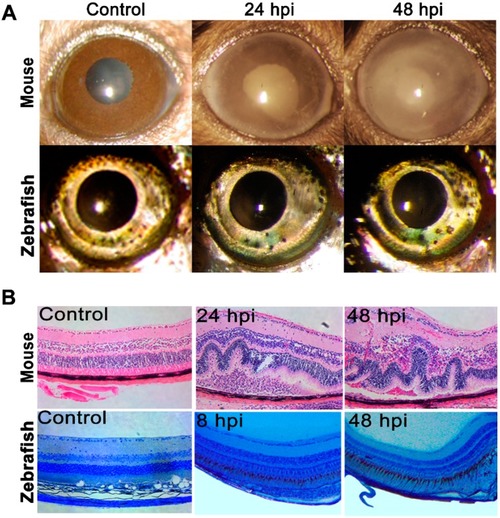
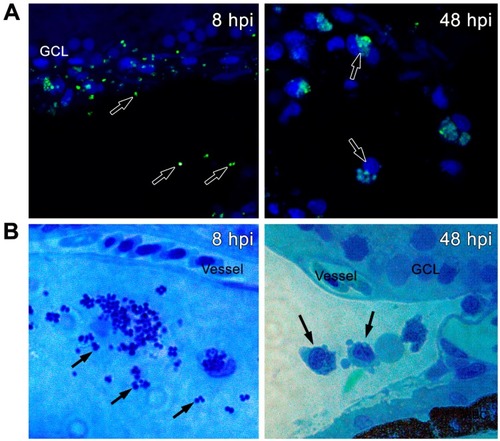
Endophthalmitis was induced through intravitreal injection of |
Endophthalmitis was induced by intravitreal injection of |
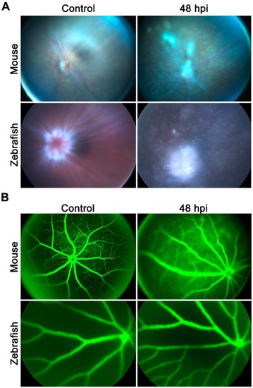
Zebrafish eyes ( |
Zebrafish eyes ( PHENOTYPE:
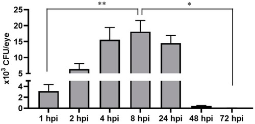
|
Zebrafish eyes ( |
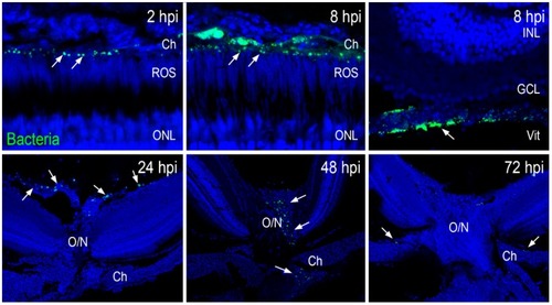
Zebrafish eyes ( |