IMAGE

Figure 4

ID
ZDB-IMAGE-200228-25
Source
Figures for Mei et al., 2019
Image
Figure Caption

Figure 4

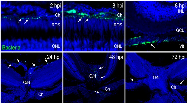
Zebrafish eyes (n = 10) were infected with GFP-positive S. aureus (250,000 CFU/eye). At indicated time points, retinal cryosections were immunostained for GFP (green) and colabeled with TO-PRO 3 for nuclei (blue). S. aureus (shown by white arrows) was initially seen in choroidal and vitreous vasculature, with the remaining bacteria being cleared through the optic nerve head with time. INL: inner nuclear layer; GCL: ganglion cell layer; Vit: vitreous chamber; O/N: optic nerve; Ch: choroid; ROS: rod outer segment.

Figure Data
Acknowledgments
This image is the copyrighted work of the attributed author or publisher, and ZFIN has permission only to display this image to its users. Additional permissions should be obtained from the applicable author or publisher of the image. Full text @ Pathogens