Differences in phagocytic efficiency of A. fumigatus and A. niger conudia at 7 hours post infection. (A) Schematic drawing of the embryo and the area imaged in the figure. (B,C) Single confocal microscopy planes of the hindbrain of zebrafish embryos injected with 150 conidia of A. fumigatus (B) and A. niger (C) at 7 hours post infection (HPI). Macrophages and neutrophils are labelled with farnesylated - membrane bound - mCherry and cytosolic eGFP respectively, and Aspergillus conidia with non membrane bound mCherry. Blow-up images without brightfield are shown below to illustrate the different appearance of phagocytosed (marked with asterisks) and free conidia (marked with hashtags). (D) Quantification of the percentage of the challenge dose left unphagocytized at 7 HPI in A. fumigatus (1,2%) versus A. niger (7,3%). Bars represent mean ± s.e.m. combined from 3 biological replicates, n = 40–47, ****p ≤ 0,0001 by t-test with Welch’s correction.

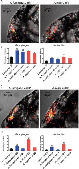
Quantification of leukocyte migration to the hindbrain at early and later stages of infection. (A,C) Representative confocal stacks of Tg (mpeg1:mCherry; mpx:GFP) zebrafish embryos with macrophages and neutrophils labelled in red and green respectively, injected with 150 conidia of A. fumigatus and A. niger at 7 HPI (A) and 24 HPI. (C) Stacks formed the basis of quantification of leukocyte migration to the hindbrain. As Macrophages and conidia were labelled with the same fluorophore, intensity of signal and morphology was used to distinguish conidia from macrophages. (B,D) Quantification of leukocyte migration in the hindbrain of embryos injected with A. fumigatus or A. niger, live or heat-killed (HK) conidia, compared to control injections (2% PVP/PBS) at 7 HPI (B) and 24 HPI. (D) *p ≤ 0,05; **p ≤ 0,01; ***p ≤ 0,001; ****p ≤ 0,0001 by one-way ANOVA with Dunnett’s multiple comparisons test, n = 21–30 per group from 3 biological replications. The quantifications represent a part of a larger data-set which is presented in full in Supplemental Fig. 1. The statistical analysis presented is based on the full dataset.

Confocal microscopy captures different infectious development of A. niger and A. fumigatus. (A) Schematic drawing of the embryo and the area imaged in the figure. (B) Still images from confocal time-lapse microscopy (Supplementary Movie SM1), depicting the typically rapid development of A. niger infection. Images from 5, 10 and 15 HPI of 150 conidia of A. niger in Tg(mpeg1:EGFP). At 15 HPI several conidia has germinated and progressive hyphal growth is evident. (C) Digitally magnified still images from Supplementary Movie SM1, aiming to capture the events of extracellular germination and hyphal growth of two separate conidia from approximately 9 HPI to 13 HPI. (D) Still images from confocal time-lapse microscopy (Supplementary Movie SM3), depicting the development of A. fumigatus infection at 5, 10 and 15 HPI, after injection of 150 conidia of A. fumigatus in Tg(mpeg1:EGFP). No germination events were detectable at the end of the time-lapse (Supplementary Movie SM3). (E) High magnification confocal microscopy showing a cluster of A. fumigatus conidia in the hindbrain of a Tg(mpeg1:EGFP) zebrafish embryo acquired at 30 hours post infection of 150 conidia. In the left panel, showing only the red fluorescence channel, several hyphae could be seen protruding from the cluster of several conidia (arrowheads). In the central panel, showing the green channel the macrophage can be seen. In the right panel, overlaying the green fluorescence channel it was clear that the cluster of conidia had been phagocytized and were contained within a macrophage. One growing hyphae could be seen to stretch the membrane of the macrophage (arrowhead). (B) White box insert in 10 HPI image represents the magnified area in image panel (C).

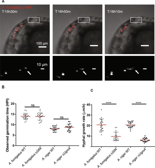
Timelapse microscopy based in vivo analysis of germination and growth rate phenotypes in galctofuranose deficient Aspergillus mutant strains. (A) Example of time-lapse microscopic assessment of hyphal growth-rate after injection of 150 conidia of the A. fumigatus ∆glfA mutant, in a pu.1 morphant embryo. Upper panels show bright field images overlaid with the red fluorescent channel. The lower panel shows a blow-up of a specific area, marked in the upper panels, from the red fluorescent channel with signal in white. Individual confocal stacks of parallel time lapse acquisitions were analyzed to estimate hyphal growth rates. (See also Supplementary Movies SM1013). (B) Analysis of germination time in confocal stacks of time-lapse microscopy experiment comparing A. fumigatus and A. niger WT to their respective Galf deficient mutant strains at an infectious burden of approximately 150 conidia. No statistically significant difference could be detected between WT and galf deficient strains in either species. (C) Analysis of approximate hyphal growth rate from confocal stacks. Plots derived from 17–21 independent hyphal growth measurements in 7–8 different pu.1 morphant embryos from three independent biological replications each. The galactofuranose deficient mutants of both Aspergillus species exhibit significantly reduced rates of hyphal elongation in vivo. (B,C) Each scatterplot depicts the 17–21 data points generated from 7–8 individual pu.1 morphant embryos covering three biological replications of each species and genotype. ****p ≤ 0,0001 by unpaired t test with Welsh correction.

Acknowledgments
This image is the copyrighted work of the attributed author or publisher, and ZFIN has permission only to display this image to its users. Additional permissions should be obtained from the applicable author or publisher of the image. Full text @ Sci. Rep.