IMAGE

Figure 2

ID
ZDB-IMAGE-191230-672
Source
Figures for Koch et al., 2019
Image
Figure Caption

Figure 2

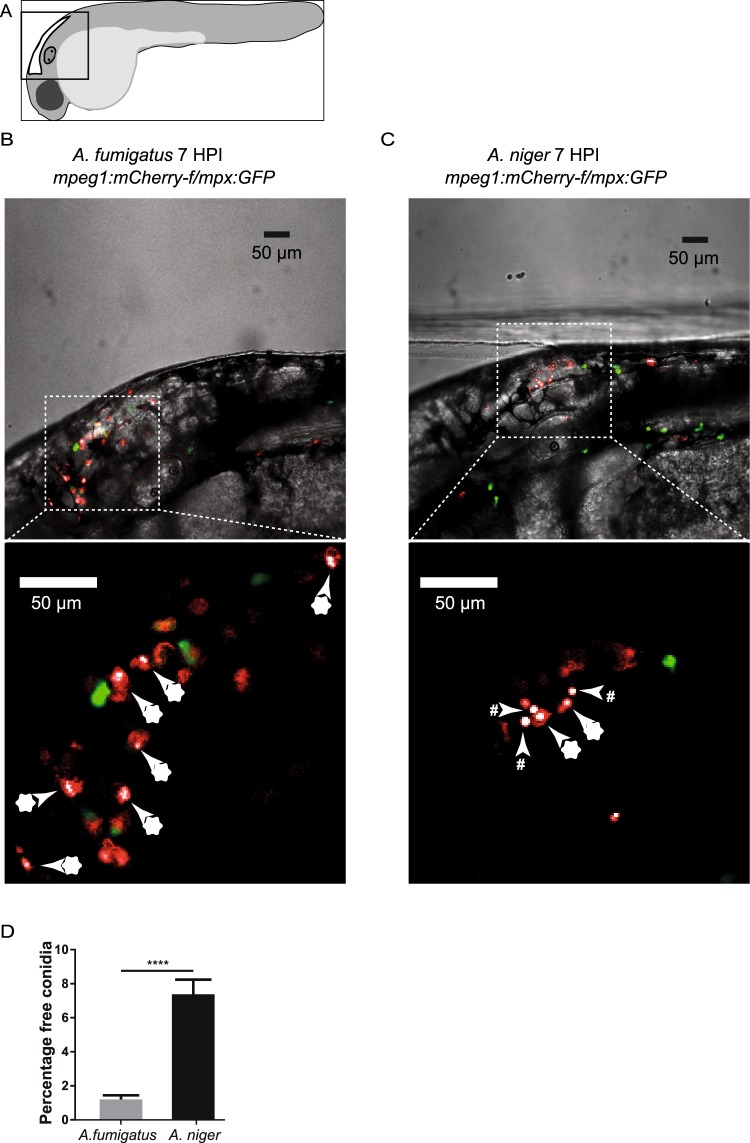
Differences in phagocytic efficiency of A. fumigatus and A. niger conudia at 7 hours post infection. (A) Schematic drawing of the embryo and the area imaged in the figure. (B,C) Single confocal microscopy planes of the hindbrain of zebrafish embryos injected with 150 conidia of A. fumigatus (B) and A. niger (C) at 7 hours post infection (HPI). Macrophages and neutrophils are labelled with farnesylated - membrane bound - mCherry and cytosolic eGFP respectively, and Aspergillus conidia with non membrane bound mCherry. Blow-up images without brightfield are shown below to illustrate the different appearance of phagocytosed (marked with asterisks) and free conidia (marked with hashtags). (D) Quantification of the percentage of the challenge dose left unphagocytized at 7 HPI in A. fumigatus (1,2%) versus A. niger (7,3%). Bars represent mean ± s.e.m. combined from 3 biological replicates, n = 40–47, ****p ≤ 0,0001 by t-test with Welch’s correction.

Acknowledgments
This image is the copyrighted work of the attributed author or publisher, and ZFIN has permission only to display this image to its users. Additional permissions should be obtained from the applicable author or publisher of the image. Full text @ Sci. Rep.