
- Title
-
Targeted sequencing of candidate genes of dyslipidemia in Punjabi Sikhs: Population-specific rare variants in GCKR promote ectopic fat deposition
- Authors
- Sanghera, D.K., Hopkins, R., Malone-Perez, M.W., Bejar, C., Tan, C., Mussa, H., Whitby, P., Fowler, B., Rao, C.V., Fung, K.A., Lightfoot, S., Frazer, J.K.
- Source
- Full text @ PLoS One
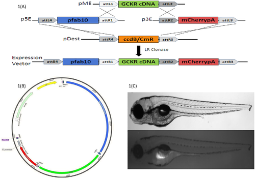
A-C. Fig 1A Multisite Gateway-based plasmid construction using Tol2 system; Fig 1B Bacterial plasmid construct (p3E-2A-mCherrypA); Fig 1C larvae (4 day post fertilization) of wild type and transgenic fish showing hepatic expression (fluorescence) after successful transgenesis of human GCKR with three disruptive mutations.
Construct:
Tg(fabp10a:Hsa.GCKR-2A-mCherry)
|

A-C. Fig 5A. Zebrafish liver histology of Hematoxylin and Eosin stained sections showing cellular differences in normal and high fat feeding for 2 weeks. A. A wild-type Tab-5 liver with normal hepatic cells and HFD showing normal liver with scattered fat cells (white arrow) showing 3+ fat without apoptosis. Fig 5B. Transgenic normal GCKR with normal hepatocytes in larvae fed on normal diet and normal liver with scattered fat cells (white arrow) in transgenic larvae fed on HFD with no apoptosis. Fig 5C. Transgenic mutant GCKR with normal diet shows liver with fatty metamorphosis and scattered hepatic cell apoptosis (white arrow) with 4+ fat. Transgenic mutant GCKR with HFD shows 4+ fat with severe cellular damage and higher fatty infiltration than the normal and transgenic normal fish. Most hepatic cells also contain vacuoles of fat and there is severe disorganization of the hepatic structure. PHENOTYPE:
|
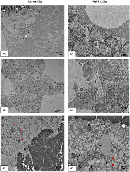
A-C. Transmission Electron Microscopy of zebrafish liver sections at medium magnification show the presence of neutral fat with HFD in normal Tab-5 Fig 6A, transgenic with normal GCKR Fig 6B, and transgenic with mutant GCKR Fig 6C. Neutral fat appears as small round vesicles with no structure. There is abnormal fat accumulation in the transgenic mutant fish with HFD (Fig 6C). Black arrows point to round vesicle like structures with empty content which are fat droplets. The red arrows point to two huge abnormal structures which appear like fusion of large neutral fat vesicles in the transgenic mutant with HFD. However, the transgenic mutant with normal diet shows abnormal structures with possible accumulation of phospholipids (red arrows) in hepatic cells suggesting abnormal metabolic function. PHENOTYPE:
|

A-C. Additional supplemental figures of WT and transgenic zebrafish livers.General observation of zebrafish larvae from three groups fed on a normal and high fat diet at 4X magnification. |