
- Title
-
Vitamin D Receptor Signaling Regulates Craniofacial Cartilage Development in Zebrafish
- Authors
- Kwon, H.J.
- Source
- Full text @ J Dev Biol
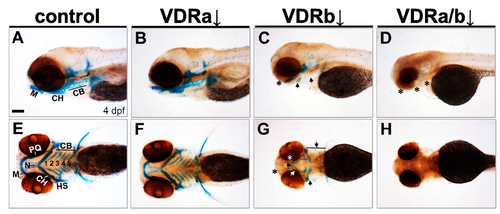
Effects of knockdown of vdr on the craniofacial cartilage development. Craniofacial cartilages were stained with Alcian blue in 4 days post-fertilization (dpf) larvae. (A,E) Wild-type controls. (B,F) vdra morphants. (C,G) vdrb morphants. (D,H) vdra/b morphants. Close-up of head lateral (A–D) and ventral (E–H) views are shown. Larvae injected with vdrb MO display absence, reduction, and malformations of cartilage components compared to controls or vdramorphants. These phenotypes are completely penetrant and consistent in vdrb morphants. The double knockdown of both vdra and vdrb leads to more severe defects, complete loss of cartilage. n ≥ 10 embryos/condition. Scale bars = 100 μm. Arrows point to reduced or malformed cartilage elements and asterisks denote missing cartilages. M, Meckel’s cartilage; PQ, palatoquadrate; CH, ceratohyal; CB, ceratobranchial; N, neurocranium; HS, hyosymplectic.
PHENOTYPE:
|
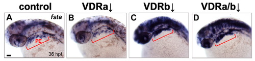
Expression of fsta in vdr morphants. (A) Wild-type control. (B) vdra morphant. (C) vdrb morphant. (D) vdra/b morphant. Expression of fsta in pharyngeal arches is upregulated in vdrb and vdra/b morphants. Upregulation throughout much of the brain, eye, and anterior spinal cord was also noticed in the embryos lacking vdrb. Images show dorsolateral views with anterior to the left at 36 hours post-fertilization (hpf). Red brackets indicate the domain of pharyngeal endoderm (PE). Scale bars = 50 μm.
|
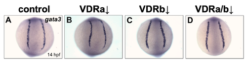
Expression of gata3 in vdr morphants. (A) Wild-type control. (B) vdra morphant. (C) vdrb morphant. (D) vdra/b morphant. Images show posterior views at 14 hpf.
|