
- Title
-
A High-Throughput Assay for Congenital and Age-Related Eye Diseases in Zebrafish
- Authors
- Brastrom, L.K., Scott, C.A., Dawson, D.V., Slusarski, D.C.
- Source
- Full text @ Biomedicines

Utilizing the eyeless masterblind (mbl) mutant as a negative control for VIZN and OMR. (A) 6 days post-fertilization control (top) and mbl−/−mutants (bottom); (B) Hematoxylin and eosin staining larvae in (A). Control larvae display normal optic structures and retinal lamination while homozygous mbl−/− mutants lack eyes; (C) Activity profile of control and, (D) mbl−/−; (E) VIZN analysis between control (n = 39) and mbl−/− (n = 28) (Mann–Whitney, p-value **** < 0.0001); (F) OMR analysis of larvae plotted as a bar graph which shows the shifts in the population between the initial and final positions (Bowker’s test of symmetry, p-value *** = 0.0004; Wilcoxon–Mann–Whitney, p-value ** = 0.0019); (G) the post-stimulus analysis which takes the difference between the final and initial position to show positional changes in individual fish. The same larvae were used for all assays. Scale bars in (B): 0.05 mm. n.s., not significant. PHENOTYPE:
|
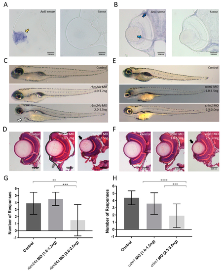
Analysis of candidate genes. Whole mount in situ hybridization on 3 dpf retinal sections for (A) rbm24a; (B) and crim1; (C) Phenotypes of control, rbm24a AUG MO at 1.0–1.2 ng, and rbm24a AUG MO at 2.0–2.5 ng; (D) H and E staining of control, rbm24a AUG MO at 1.0–1.2 ng, and rbm24a AUG MO at 2.0–2.5 ng; (E) Phenotypes of control, crim1 AUG MO at 1.0–1.5 ng, and crim1 AUG MO at 2.5–3.0 ng; Same magnification as (A); (F) H and E staining of control, crim1 AUG MO at 1.0–1.5 ng, and crim1 AUG MO at 2.5–3.0 ng. Same magnification as (B); (G) VIZN analysis of control (n = 19), rbm24a AUG MO at 1.0–1.2 ng (n = 18), and rbm24a AUG MO at 2.0–2.5 ng (n = 10) (Mann–Whitney p-value ** = 0.0068, *** = 0.0003); (H) VIZN analysis of control (n = 39), crim1 AUG MO at 1.0–1.5 ng (n = 31), and crim1 AUG MO at 2.5–3.0 ng (n = 18) (Mann–Whitney p-value **** < 0.0001). Yellow arrow indicates lens staining. Light blue arrow denotes ganglion cell layer staining. Dark blue arrow indicates choroid staining. Black arrows point to microphthalmic eyes. White arrow indicates cardiac edema. Scale bars in (A) and (B): 0.04 mm, and in (D) and (F): 0.05 mm. EXPRESSION / LABELING:
PHENOTYPE:
|
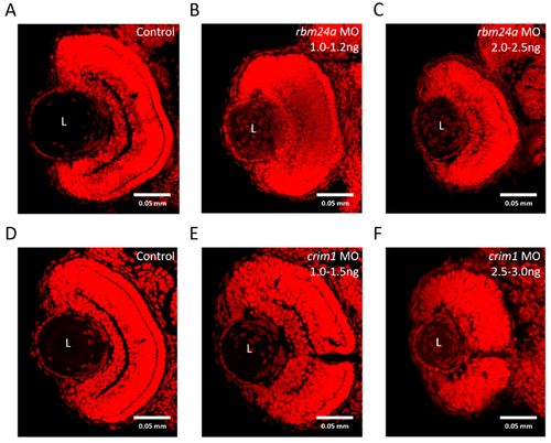
rbm24a and crim1 morphants have congenital eye defects. (A) Control; (B) rbm24a AUG MO 1.0–1.2 ng; (C) rbm24a AUG MO 2.0–2.5 ng; (D) control; (E) crim1 AUG MO 1.0–1.5 ng, and (F) crim1 AUG MO 2.5–3.0 ng. L denotes the lens. Scale: 0.05 mm. TOPRO3-stained nuclei appear red. PHENOTYPE:
|