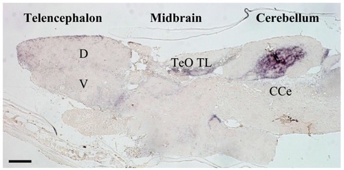
Photomicrograph of mineralocorticoid receptor (MR) in-situ hybridization in medaka brain. MR expression is restricted to a number of important areas that likely correspond to homologous brain regions containing MR in other vertebrates including those of humans [35]. Telencephalic regions exhibiting higher MR expression include the ventral parts of the lateral zone of the dorsal telencephalon (putative fish homologue of the mammalian hippocampus [36]), and commissural and subcommissural nuclei of the telencephalon (V; putative fish homologue to the mammalian amygdala) [36]. In the diencephalon, the hypothalamic preoptic area, inferior lobe of the hypothalamus, and glomerulus complex of the thalamus exhibit MR expression, as do the mesencephalic tegmentum and granular layer of the optic tectum. MR expressed markedly in some regions of the cerebellum. CCe, corpus cerebelli; D, dorsal telencephalic area; TeO, tectum opticum; TL, torus longitudinalis; V, ventral telencephalic area. The detailed expression profiles are described in Sakamoto et al. [13]. Ubiquitously expressed GR in the brains of rodent and fish, as well as MR expression in the rodent brain, have been illustrated elsewhere [15,37,38]. Scale bar: 100 μm.

Acknowledgments
This image is the copyrighted work of the attributed author or publisher, and ZFIN has permission only to display this image to its users. Additional permissions should be obtained from the applicable author or publisher of the image. Full text @ Nutrients