
- Title
-
CXCR4 signaling regulates metastatic onset by controlling neutrophil motility and response to malignant cells
- Authors
- Tulotta, C., Stefanescu, C., Chen, Q., Torraca, V., Meijer, A.H., Snaar-Jagalska, B.E.
- Source
- Full text @ Sci. Rep.
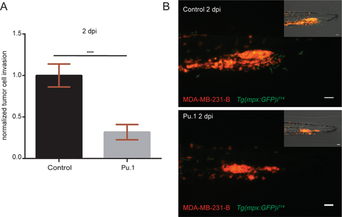
Myeloid cell depletion impairs tumor cell invasion. (A) Relative tumor invasion was compared at 2 dpi in Pu.1 morphants, depleted of neutrophils and macrophages, and larvae injected with control morpholino (68% inhibition). Two-tailed un-paired t-test with Welch’s correction (****p < 0.0001) was performed on a pool of two biological replicates (Control: n = 84, Pu.1: n = 67). Data are mean ± SEM. (B) Top panel shows MDA-MB-231-B cells forming a tumor mass and invading the tail fin tissue (bright field image, top right), while surrounded by GFP expressing neutrophils in 2 dpi Tg(mpx:GFP)i114 injected with a control morpholino. In the bottom image, neutrophils are absent due to Pu.1 knockdown and a smaller tumor mass is formed compared to the control condition, resulting in impaired invasion of the local tissue (bright field, top right). Scale bar: 50 µm. Micrographs were acquired using a Leica MZ16FA fluorescent microscope coupled to a DFC420C camera.
|
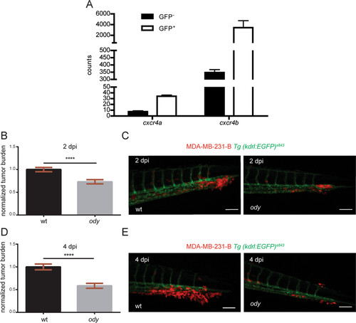
cxcr4b is highly expressed in neutrophils and loss of function results in reduced triple negative breast cancer burden. (A) cxcr4a and cxcr4b expression levels were quantified in neutrophils and compared to the GFP negative cell population. Data are read counts from RNA sequencing performed on three biological replicates. FACS-sorted neutrophils were obtained from 5 dpf Tg(mpx:GFP)i114 larvae. cxcr4a and cxcr4b gene expression was enriched in neutrophils compared to GFP negative cells in zebrafish larvae (~4-fold and ~10-fold, respectively). cxcr4b was highly expressed in neutrophils compared to cxcr4a(~100-fold increased gene expression). (B) Relative metastatic tumor burden of MDA-MB-231-B-DsRed cells was quantified in ody and wt siblings at 2 dpi. Data are mean ± SEM of two independent experiments (wt: n = 64, ody: n = 57). Un-paired t-test ****p < 0.0001. (C) MDA-MB-231-B tumor cells established a secondary tumor mass, with initiation of single cell extravasation, in wt larvae, whereas a phenotype inhibition was found in ody mutants at 2 dpi (22.5% reduction). (D) MDA-MB-231-B tumor burden was measured in wt and cxcr4b null mutants at 4 dpi, at the metastatic site where secondary growth began at 2 dpi. A 40.5% reduction in tumor burden was observed. Data are mean ± SEM of two independent experiments (wt: n = 59, ody: n = 43). Un-paired t-test, with Welch’s correction ****p < 0.0001. (E) Highly invasive cancer cells displayed aggressive and metastatic features in wt siblings, whereas few cells remained in the CHT region of 4 dpi odylarvae. Scale bars: 50 µm. Micrographs are acquired using a Leica MZ16FA fluorescent microscope coupled to a DFC420C camera.
|
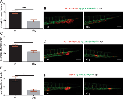
cxcr4b deficient host blocks metastatic burden of different tumor types. (A) Metastatic burden was assessed in 4 dpi zebrafish larvae engrafted with the triple negative breast line MDA-MB-157 mCherry. A 52% reduction was found. Data are mean ± SEM of two independent experiments (wt: n = 42, ody: n = 28). Un-paired t-test, with Welch’s correction ****p < 0.0001. (B) Secondary tumor mass, extravasation and invasion failed to occur in ody mutants compared to wt siblings. (C) A significantly lower tumor burden in cxcr4b deficient larvae was observed when the prostate cancer PC3-M-Pro4-Luc2 mCherry or td-tomato cell line was implanted (38% reduction). Data are mean ± SEM of two independent experiments (wt: n = 48, ody: n = 46). Un-paired t-test ****p < 0.0001. (D) Prostate cancer early metastasis formation, characterized by a solid tumor mass formation in the CHT region of zebrafish larvae, occurred in wt siblings and was significantly decreased when Cxcr4b signaling was impaired in the host. (E) Relative metastatic burden of Ewing sarcoma cell line WE-68 td-tomato was affected in ody mutants compared to wt larvae at 4 dpi (70% reduction in tumor burden in the tail fin). Data are mean ± SEM of two independent experiments (wt: n = 69, ody: n = 39). Un-paired t-test, with Welch’s correction ****p < 0.0001. (F) Ewing sarcoma cells formed a compact and expanding tumor mass in the CHT region, between the dorsal aorta and the caudal vein. A reduced tumor cell aggregate was present in the ody mutant line at 4 dpi. Scale bars: 50 µm. Micrographs were acquired using a Leica MZ16FA fluorescent microscope coupled to a DFC420C camera. |
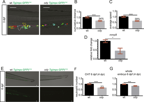
cxcr4b deficiency affects neutrophil physiological motility and development. (A) Neutrophil movement was recorded for 30 minutes and tracks showed reduced motility in ody compared to wt siblings in the tail fin region where tumor metastasis formation generally takes place. Scale bars: 50 µm. Time-lapse microscopy was performed using a Leica TCS SPE confocal microscope with a HC APO 20x DRY objective (0.7 N.A.). Neutrophil motility was assessed in wt and ody larvae at 3 dpf, measuring cumulative distance (B) and average speed (C) of each phagocyte, localized in the CHT region. (B) Un-paired t-test **** p < 0.0001 and (C) Un-paired t-test, with Welch’s correction ****p < 0.0001. Data are mean ± SEM of two independent experiments and values were calculated from 54 tracks (wt: n = 7) and 58 tracks (ody: n = 8). (D) mmp9 expression in 6 dpf ody and wt siblings. *p = 0.02, unpaired t-test. (E,F) Number of neutrophils in wt and ody in the CHT region at 6 dpf is shown. A lower neutrophil number was found in the CHT region in cxcr4b −/− larvae (32% reduction), as shown by top and bottom micrographs (E) and quantified in (F). Un-paired t-test **** p < 0.0001. Data are mean ± SEM of two independent experiments (wt: n = 35, ody: n = 36). (G) A significant reduction in total neutrophil number was found in ody larvae at 6 dpf. In (G) wt: n = 35, ody: n = 36. Data are mean ± SEM (pool of two independent experiments). Un-paired t-test with Welch’s correction ***p = 0.0007. |
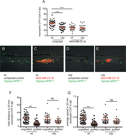
cxcr4b loss of function influences neutrophil response to cancer cells initiating early metastases. (A) Neutrophil response to metastatic cancer cells was assessed by measuring neutrophil number in the CHT in wt and ody larvae at 4 dpi (6 dpf). In control conditions, neutrophils left the CHT when tumor cells were present, whereas they failed to respond, remaining in the CHT, in ody mutants (B). Kruskal-Wallis, with Dunn post hoc test **** p < 0.0001 (number of uninjected embryos is the same as in graph in Fig. 4F; number of engrafted embryos is wt: n = 29 and ody: n = 25). Images were acquired using Leica MZ16FA fluorescent microscope coupled to a DFC420C camera. Scale bars: 50 µm. (B,C) Neutrophil tracking in 6 dpf larvae showed stationary behavior of neutrophils in the presence of tumor cells in wt siblings compared to uninjected controls. (D,E) Neutrophils maintained the same migratory behavior in odymutants, in presence of MDA-MB-231-B and in uninjected larvae. (F,G) Neutrophil motility was quantified for 30 minutes, measuring total distance and average speed of each neutrophil in the CHT region. Data are mean ± SD (uninjected wt: n = 46 tracks from 7 larvae; MDA-MB-231-B wt: n = 32 tracks from 5 larvae; uninjected ody: n = 37 tracks from 7 larvae; MDA-MB-231-B ody: n = 27 tracks from 5 larvae). One-way ANOVA, with Bonferroni post-hoc test. |