
- Title
-
Propyl gallate inhibits hepatocellular carcinoma cell growth through the induction of ROS and the activation of autophagy
- Authors
- Wei, P.L., Huang, C.Y., Chang, Y.J.
- Source
- Full text @ PLoS One
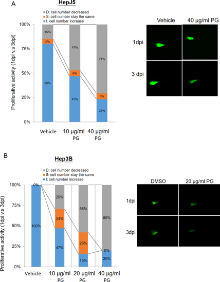
PG suppresses cell proliferation in a zebrafish model. The xenotransplantation assay was performed using zebrafish to determine the efficacy of PG treatment in HCC. Hep3B and HepJ5 cells were implanted into the embryo yolk and then exposed to different doses of PG (0–40 μg/ml). The proliferative activity in the HCC cell lines was compared by monitoring the fluorescence intensity on day 1 and day 3 post-injection (1 dpi and 3 dpi) of PG. (A) PG at concentrations of 10 μg/ml and 40 μg/ml reduced the cell number increase in the embryo population (from 80% vehicle to 47% and 24%, respectively) in HepJ5 cells. A decreased fluorescence intensity was shown after 3 days in HepJ5 cells with 40 μg/ml PG treatment. (B) In Hep3B cell lines, the cell number increase in the embryo population was decreased from 100% (vehicle) to 47%, 16% and 20% (10 μg/ml, 20 μg/ml, and 40 μg/ml, respectively). Treatment with 20 μg/ml PG dramatically decreased the fluorescence intensity in Hep3B cells compared with vehicle. |