
- Title
-
The Ancient Origins of Neural Substrates for Land Walking
- Authors
- Jung, H., Baek, M., D'Elia, K.P., Boisvert, C., Currie, P.D., Tay, B.H., Venkatesh, B., Brown, S.M., Heguy, A., Schoppik, D., Dasen, J.S.
- Source
- Full text @ Cell
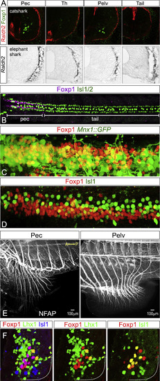
Development of Fin MNs in Danio rerio, Scyliorhinus canicula, and Callorhinchus milii, Related to figure 3 (A) Raldh2 expression is not detected in fin-level MNs of catshark (Scyliorhinus canicula) or elephant shark (Callorhinchus milii). In both species, expression of Raldh2 is detected in the tissue surrounding the spinal cord, similar to the pattern observed in mice. (B) Dorsal view of 48hpf zebrafish embryo showing Foxp1 expression at pectoral (pec) levels. Isl1 staining labels a subset of MNs as well as larger diameter sensory neurons. Foxp1 expression in the ventral spinal cord extends from the hindbrain to the rostral spinal cord, adjacent to myotomes m2-m5, consistent with previous studies (Ma et al., 2010). (C and D) High-magnification images of Foxp1 protein expression at pectoral levels in zebrafish, showing colocalization with Mnx1::GFP (at 72 hpf) and Isl1 (at 48 hpf). In zebrafish a subset of Foxp1+ neurons express Isl1 at relatively weak levels. (E) Whole-mount NFAP staining showing projection of motor axons toward the pectoral and pelvic fin in catshark. (F) Sections of catshark embryos showing a subset of Foxp1+ MNs express Isl1 or Lhx1. In catshark LMCm and LMCl neurons appear to be intermixed. |
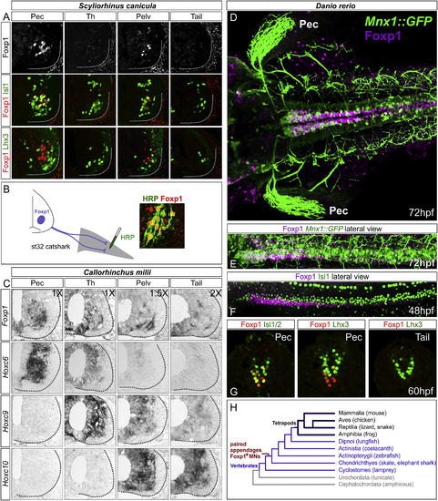
LMC Neurons Are Present in Multiple Species of Aquatic Vertebrates (A) Expression of MN determinants in the small-spotted catshark, Scyliorhinus canicula, at st31. Foxp1+ MNs are detected at fin levels but are absent in inter-fin (“Th”) and tail levels. Lhx3+ MMC-like neurons are present at all levels and lack Foxp1. (B) Retrograde labeling of MNs after HRP injection into the pectoral fin in Scyliorhinus. Foxp1+ MNs co-label with HRP. (C) In situ hybridization showing expression of MN markers and Hox genes in Callorhinchus milii at st31. Foxp1 is detected at fin levels and also at inter-fin and tail levels, possibly reflecting spinal INs. Hoxc6 is expressed at pectoral levels, Hoxc9 at inter-fin levels, and Hoxc10 at pelvic fin levels. Differences in relative image magnification are shown. (D–G) Expression of MN determinants in zebrafish. (D) Dorsal view of a 72-hpf Mnx1::GFP embryo showing expression of Foxp1 at pectoral levels. GFP also labels projections to the pectoral (Pec) fin. Foxp1 is expressed in INs at more caudal segments. (E) Lateral view showing coexpression of GFP in a subset of Foxp1+neurons. (F) Lateral view of 48-hpf embryo showing Foxp1 and Isl1 expression. (G) Transverse sections showing that Foxp1+ neurons lack Lhx3. (H) Simplified chordate phylogeny indicating the presence of a limb/fin innervation program in the common ancestor to skates and tetrapods. |
Reprinted from Cell, 172, Jung, H., Baek, M., D'Elia, K.P., Boisvert, C., Currie, P.D., Tay, B.H., Venkatesh, B., Brown, S.M., Heguy, A., Schoppik, D., Dasen, J.S., The Ancient Origins of Neural Substrates for Land Walking, 667-682.e15, Copyright (2018) with permission from Elsevier. Full text @ Cell