
- Title
-
Color Processing in Zebrafish Retina
- Authors
- Meier, A., Nelson, R., Connaughton, V.P.
- Source
- Full text @ Front. Cell. Neurosci.
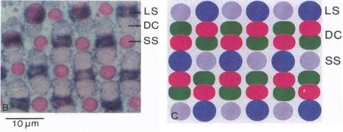
Zebrafish cone mosaic. In adult zebrafish, the cone mosaic is highly structured, with red and green double cones (DC) between blue (Long single, LS) and UV (Short single, SS) cones, with the red cone always next to the blue cone and the green cone always next to the UV cone (from Robinson et al., |
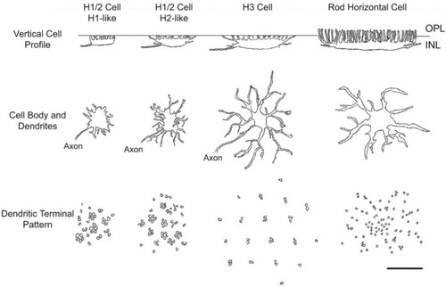
Zebrafish horizontal cells (HCs). HCs in zebrafish retina are morphologically distinct. H1-like cells are smaller in vertical profile and somal size compared to H2 and H3 cells, and all types have distinct dendritic connections with presynaptic photoreceptors (PRs). Reproduced with permission from Song et al. ( |
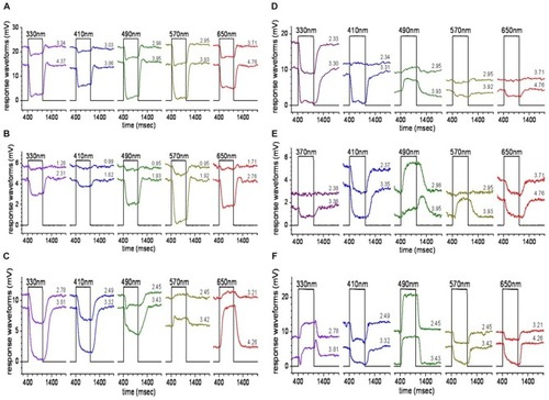
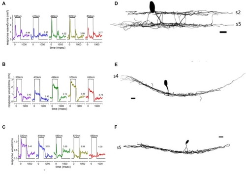
HC responses. Stimulation of zebrafish eyecups with light of different spectral wavelengths evokes different responses from zebrafish HCs. |
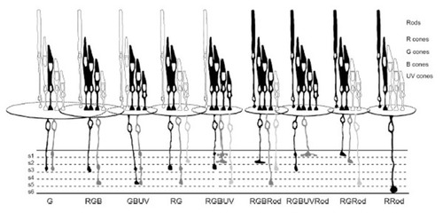
Bipolar cells make diverse connections with PRs. Summary figure (reproduced with permission from Li et al., |
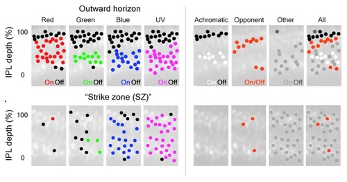
Color inputs to inner retina are segregated in the IPL. BC terminals in the larval zebrafish IPL can be segregated based on the spectral inputs they respond to across the visual scene. (Left) BC-terminal excitation by R, G, B, and UV stimuli are represented as red, green, blue and magenta dots located throughout the IPL; inhibitory responses are represented as black dots. The distribution of ON-responses varies based on the spectral stimulus and which part of the visual scene (outer horizon vs. “strike zone (SZ)”) is being viewed. The “SZ” represents looking forward and upward at prey items. (Right) Subsequent classification of the different spectral inputs from BCs into the IPL identified achromatic and color-opponent regions in this synaptic layer that were most evident outside the “SZ”. For the right panels: black dots = achromatic OFF-responses, white dots = achromatic ON-responses, red dots = color-opponent responses, gray dots = “other” (taken from Figure 3, Zimmermann et al., |
Spectral responses of ACs are associated with specific cell types. Transient ON-OFF amacrine cells (AC) responses |
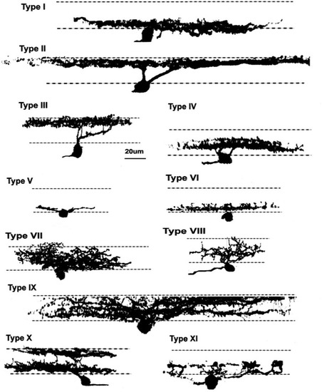
Zebrafish ganglion cells (GCs). Di-I labeling of GCs in whole mount tissue identified 11 morphological types, based on dendritic arborization patterns in the IPL. These patterns included processes restricted to 1–2 sublaminae, as well as more diffuse arborization patterns.Reproduced with permission from Mangrum et al. ( |
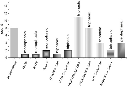
GC spectral responses. Loose patch recordings from larval zebrafish GCs revealed a diversity of response types, most of which are spectrally multiphasic. Triphasic responses were the most abundant type identified and could be subdivided into three distinct spectral patterns. Bi-, tetra-, and pentaphasic responses were also recorded (Connaughton and Nelson, |