FIGURE SUMMARY
Title

Leveraging Zebrafish to Study Retinal Degenerations

Authors
Angueyra, J.M., Kindt, K.S.
Source
Full text @ Front Cell Dev Biol

Structure of the zebrafish eye and retina. (A) Anatomy of the zebrafish eye: DIC image of a cryosection from a 6 days post-fertilization (6 dpf) larval eye highlighting the main structures of the vertebrate eye including cornea, lens, retina, and retinal pigment epithelium (RPE). (B) The larval retina is organized into highly structured layers: Overlay of a DIC image and the fluorescent nuclear marker DAPI of a cryosection from a 6 dpf larva showing the different retinal layers, including the outer nuclear layer (ONL) which contains the cell bodies of photoreceptors (rods and cones). Photoreceptors make synapses in the outer plexiform layer (OPL) with bipolar and horizontal cells. The inner nuclear layer (INL) contains the cell bodies of horizontal, bipolar and amacrine cells, while the ganglion cell layer (GCL) contains the cell bodies of retinal ganglion cells (RGC). Bipolar cells provide excitatory synaptic input to RGC in the inner plexiform layer (IPL), while amacrine cells modulate this input both pre- and post-synaptically. (C) The adult retina retains the same layered structure: Overlay of a DIC image and DAPI of a cryosection from an adult zebrafish. (D) The zebrafish retina contains 4 subtypes of cones: DIC image of a cryosection from an adult zebrafish showing the short-single or ultraviolet-wavelength sensitive cones (UV-cones), the long-single or short-wavelength sensitive cones (S-cones), and the double cones which correspond to the middle- and long-wavelength sensitive cones (M- and L-cones). (E) Mosaic arrangement of zebrafish cone photoreceptors: Confocal image of a whole-mounted retina of a double-reporter transgenic lines to identify UV-cones [Tg(sws1:GFP)kj9, magenta] and S-cones [tg(sws2:mCherry)ua3011, blue] overlayed with DAPI (gray), allowing the identification of the nuclei of M- and L-cones between the rows of UV- and S-cones. (F) Diagram of the vertebrate retina and the retinal cells. Inset highlights the synapse between cones and horizontal and bipolar cells, where the cone synaptic terminal contains synaptic vesicles (white) attached to the synaptic ribbon (black). In close apposition to the ribbon, the dendrites on On-bipolar cells (On-BCs) invaginate into the synaptic terminal and are flanked by two horizontal cell (HC) processes. Off-bipolar cells make more basal contacts in close proximity but not apposed to the synaptic ribbon.

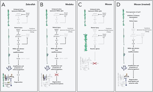
Pathways to retinal regeneration. (A) Zebrafish are able to completely regenerate their retina via Müller glia. In the uninjured retina, Müller glia is kept quiescent by inhibiting the expression of the genes that control regeneration through Notch signaling and repression by the transcription factor Six3b and the microRNA let7, amongst others. During retinal injury, cell death and inflammation lead to the release of cytokines and growth factors (especially TNFα), which activate receptors and kinases in the Müller glia, leading to activation of β-catenin and phosphorylation of the transcription factor Stat3, which in turns leads to the production of the transcription factor ascl1 and lin28 microRNA, and the activation of the genes that control Müller glia division and dedifferentiation. lin28 also inhibits the production of let7, releasing the inhibition of this pathway. Expression of the transcription factor Pax6b (normally inhibited by the microRNA mi203), allows the amplification and production of retinal progenitors which are then able to redifferentiate into any retinal neurons or Müller glial cells. (B) In medaka fish, lack of production of Sox2 after the production of retinal progenitors restricts their fate to photoreceptors and does not allow the production of other cell types, including new Müller glia. (C) In mice and other mammals, retinal injury does not lead to expression of ascl1 and the rest of regeneration-related genes. Instead Müller glia activate the production of intermediate filaments and increase their size (reactive gliosis) and do not lead to the production of new retinal cells or injury repair. (D) Retinal regeneration can be stimulated in mice by artificially overexpressing ascl1 and inhibiting epigenetic changes (histone deacetylation in particular). With this treatment, Müller glial cells are able to divide and produce retinal progenitors, but their fate is restricted to bipolar and amacrine cells.

Tools to study retinal circuits. (A) Confocal image of the UV-cone reporter line Tg(sws1:GFP)kj9 retina (magenta), overlaid with DAPI nuclear staining (cyan) and a transmitted DIC image. (B) Confocal image of the S-cone reporter line Tg(-3.5opn1sw2:EGFP)k11 retina (blue), overlaid with DAPI nuclear staining (cyan) and a transmitted DIC image. (C) Bipolar cell labeling using PKCα immunolabeling (yellow) and the On-bipolar reporter line Tg(grm6b:EGFP)zh1 (blue). Rod-contacting bipolar cells are brightly labeled by the PKCα antibody, while another subset of bipolars is more dimly labeled. Some of these are doubly labeled with the transgenic line. (D) Immunolabeling of off-bipolar synapses with photoreceptors using an antibody against the inotropic glutamate receptor type 4 (gria4), in the background of a double transgenic reporter line Tg(sws1:GFP)kj9 (magenta) and Tg(grm6b:EGFP)zh1 (blue). Inset shows that the punctate labeling in the IPL overlaps with the synaptic terminals of cones and the bipolar cell dendrites. (E) Sparse labeling of horizontal cells using DiI (yellow) in the background of the S-cone reporter line Tg(-3.5opn1sw2:EGFP)k11. Image corresponds to a maximal intensity projection of a confocal stack, where synaptic contacts between the horizontal cell and S-cones are apparent. (F) Maximal intensity projection of an orthogonal view restricted to the box gray in (E), highlighting the invaginations of horizontal processes into the cone synaptic terminals. This particular projection is oriented through a row of UV- and S-cones and the bigger horizontal processes most likely correspond to invaginations into UV-cone terminals.

Acknowledgments
This image is the copyrighted work of the attributed author or publisher, and ZFIN has permission only to display this image to its users. Additional permissions should be obtained from the applicable author or publisher of the image. Full text @ Front Cell Dev Biol