
- Title
-
Next-generation and further transgenerational effects of bisphenol A on zebrafish reproductive tissues
- Authors
- Akhter, A., Rahaman, M., Suzuki, R.T., Murono, Y., Tokumoto, T.
- Source
- Full text @ Heliyon
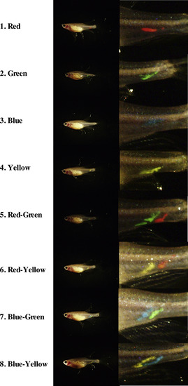
The sexually mature females and males (3-month-old fish of 0.2 ± 0.04 g in body weight) used for the treatments were selected and marked by injection of fluorescent dye (Visible Implant Elastomer Tag, Northwest Marine Technology, NW). Different 8 patterns of color were used to distinguish individuals in a group of 8 fishes. Marked fish were kept in same tank as one experimental group ( |

Adverse effects of BPA treatment on ovaries in the F1 generation of zebrafish. (A) Outside view of a normal β-roy female. (B) Parental male zebrafish were fed BPA-containing food (10 mg of BPA/g food) for 6 months. Then fish were mated with untreated fish (housed separately from experimental fish) and offspring were raised on control food. Outside view of raised female fish generated from BPA-treated males. Note that all indicated fish are seems like males in bright field but they have fluorescent oocytes in abnormal ovaries. Photographs of whole fish and ovarian tissues under bright field (B. field) and GFP filter views (GFP) are shown. |
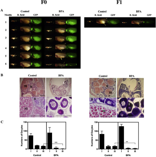
Adverse effects of BPA treatment on ovaries. (A) The morphology of ovaries of F0 females during the treatment was monitored by fluorescence observations performed at 1-month intervals (F0). Photographs of representative F1 females from the control (Control) and BPA-treated (BPA) groups were taken before histological analysis (F1). Photographs of whole fish and ovarian tissues under bright field (B. field) and GFP filter views (GFP) are shown. (B) Sections of control-treated (Control) and BPA-treated (BPA) females. Developmental stages of oocytes are indicated according to Selman et al. as follows: I (early vitellogenic oocytes, perinucleolar oocyte), II (Mid-vitellogenic oocytes) and III (fully grown oocytes) (Selman et al., 1993). The scale bars indicate 50 μm. (C) Number of oocytes in stages I, II or III in sections of control-treated (Control) and BPA-treated (BPA) females were counted in three sections prepared from three different fishes. Vertical lines indicate standard deviation. ** indicate statistically significant differences between the values for the BPA-treated group and the control group at the P < 0.01 level. |
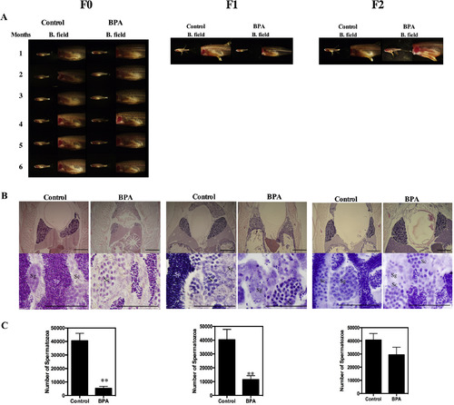
Adverse effects of BPA treatment on testes. (A) The morphology of testes during the treatment was monitored by microscopic observations performed at 1-month intervals (F0). Photographs of representative F1 and F2 males from the control (Control) and BPA-treated (BPA) groups were taken before histological analysis (F1 and F2). Photographs of whole fish and testes under bright field (B. field) are shown. (B) Sections of control (Control) and BPA-treated (BPA) males. Cysts containing spermatogonia (Sg), spermatocyte (Sc), spermatozoa (Sp) can be seen (Rupik et al., 2011). The scale bars indicate 50 μm. (C) Number of spermatozoa (Sp) in sections of control-treated (Control) and BPA-treated (BPA) males were counted in three sections prepared from three different fishes. Vertical lines indicate standard deviation. ** indicate statistically significant differences between the values for the BPA-treated group and the control group at the P < 0.01 level. |

Abnormal embryonic development induced by parental BPA treatment. The morphologies of normal embryos from control (Control) fish and abnormal embryos from BPA-treated fish in generations F1 to F3. Photographs of three representative phenotypes of abnormal embryos in each generation are shown. Most of the embryos died from coagulation before hatching. Approximately five to fifteen percent of the surviving embryos exhibited abnormalities in all generations. It was observed that several abnormalities, including skeletal spine curvature (ssc), cardiac edema (ce), problems with yolk sacreabsorption (ysr), non-detachment of the tail from the yolk (dty), and double-head (dh), occurred randomly in individuals. |