
- Title
-
Angiotropism and extravascular migratory metastasis in cutaneous and uveal melanoma progression in a zebrafish model
- Authors
- Fornabaio, G., Barnhill, R.L., Lugassy, C., Bentolila, L.A., Cassoux, N., Roman-Roman, S., Alsafadi, S., Del Bene, F.
- Source
- Full text @ Sci. Rep.

Cutaneous melanoma cells and non-malignant melanocytes show different migratory properties in zebrafish. (A), (A’) and (A”) Different images of a 3 dpi larva injected with Hermes-GFP cells, showing no melanocytes outside the yolk cavity. (B), (B’) and (B”) Different images of a 2 dpi embryo injected with C8161-GFP cells, showing numerous melanoma cells spread all over the body of the fish. Pictures were taken with a 10 × dry objective, employing a Zeiss LSM 700 confocal microscope. Scale bar is 50 µm, green shows melanocytes, red shows zebrafish blood vessels. |
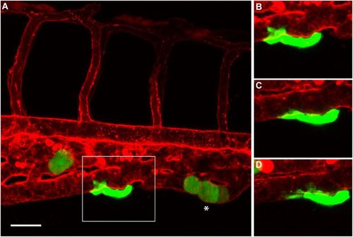
Angiotropism in zebrafish xenograft of cutaneous melanoma. (A) A larva injected with C8161-GFP cells, displaying an angiotropic cell (in the square) extending along the external surface of the caudal vein. (B–D) are time-lapse images of the same angiotropic cell taken at 0, 4 and 8 hours after the beginning of the imaging. The images were obtained employing a Zeiss LSM 700 confocal microscope (25 × oil objective), starting from 30 hours post injection. Scale bar is 20 µm, green shows cutaneous melanoma cells, white asterisk shows intravascular melanoma cells, red shows zebrafish blood vessels. PHENOTYPE:
|

Angiotropism in zebrafish xenograft of uveal melanoma. (A) A larva injected with OMM 2.3-GFP cells, displaying a micrometastasis of angiotropic cells (in the square) cuffing the external surface of an intersegmental vessel. (B–D) are time-lapse images of the same angiotropic cells taken at time 0, 4 and 8 hours after the beginning of the imaging. The images were obtained employing a Zeiss LSM 880 confocal microscope (40 × water objective), starting from 30 hours post injection. Scale bar is 20 µm, green shows melanoma cells, red shows zebrafish blood vessels, white arrows show pseudopodial protrusions formed by angiotropic cells, white asterisk shows intravascular melanoma cells, yellow arrow shows melanoma cells trapped in an intersegmental vessel. |