
Image
|
Figure Caption
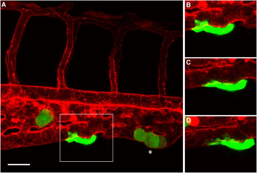
Fig. 5
Angiotropism in zebrafish xenograft of cutaneous melanoma. (A) A larva injected with C8161-GFP cells, displaying an angiotropic cell (in the square) extending along the external surface of the caudal vein. (B–D) are time-lapse images of the same angiotropic cell taken at 0, 4 and 8 hours after the beginning of the imaging. The images were obtained employing a Zeiss LSM 700 confocal microscope (25 × oil objective), starting from 30 hours post injection. Scale bar is 20 µm, green shows cutaneous melanoma cells, white asterisk shows intravascular melanoma cells, red shows zebrafish blood vessels.
Figure Data
Acknowledgments
This image is the copyrighted work of the attributed author or publisher, and
ZFIN has permission only to display this image to its users.
Additional permissions should be obtained from the applicable author or publisher of the image.
Full text @ Sci. Rep.