
- Title
-
Connexin Communication Compartments and Wound Repair in Epithelial Tissue
- Authors
- Chanson, M., Watanabe, M., O'Shaughnessy, E.M., Zoso, A., Martin, P.E.
- Source
- Full text @ Int. J. Mol. Sci.

The different phases of airway epithelium regeneration after wounding. The top left images show the histology of the pseudostratified airway epithelium after culturing human airway epithelial cells for 1.5 months on Transwell filter, and the capacity of the epithelium to repair after wounding. BC: basal cell; CC: ciliated cell; GC: goblet cell. The scheme illustrates the steps involved in airway epithelium repair after injury (blue arrows); wound closure is reached within 3–4 days. Orange triangle: CK5-expressing quiescent basal cells; orange square: CK5 and CK14-activated basal cells; light green square: early progenitor cells; dark green square: late progenitor cells. Early differentiation (passage from proliferating cells to early progenitors) is dictated in part by Notch activation (green arrows). Cell division arrest and later differentiation requires increased expression of miR-449. The relative changes in connexin expression (Cx26, Cx30, Cx31) is illustrated for the different stages of the repair process by the size of the fonts. PPARγ signalling also contributes to BC differentiation and decreases connexin expression (red arrow). Bar: 50 µm. |
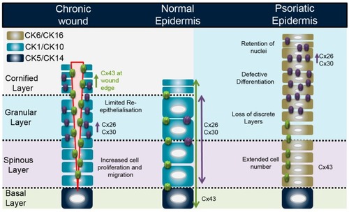
Connexin expression profile in the normal epidermis, at a chronic wound edge, and representative state in a psoriatic epidermis. |