
- Title
-
The protein tyrosine phosphatase 1B inhibitor MSI-1436 stimulates regeneration of heart and multiple other tissues
- Authors
- Smith, A.M., Maguire-Nguyen, K.K., Rando, T.A., Zasloff, M.A., Strange, K.B., Yin, V.P.
- Source
- Full text @ NPJ Regen Med
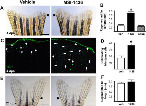
MSI-1436 stimulates adult zebrafish caudal fin regeneration. a Representative images of caudal fin regeneration 4 days post-amputation (dpa) in vehicle- and MSI-1436-treated fish. b Quantification of regenerated caudal fin length 4 dpa. Values are means ± S.E. (n = 12–16). *P < 0.01 compared to vehicle-treated fish. c Representative images of caudal fin blastemas immunostained with antibodies to phosphorylated histone 3 (H3P). Arrows show H3P-positive cells. d Quantification of H3P-positive cells 4 dpa. Values are means ± S.E. (n = 6–8). *P < 0.01 compared to vehicle-treated fish. e Representative images of caudal fin morphology after regeneration is complete at 21 dpa. f Quantification of regenerated caudal fin length 21 dpa. Values are means ± S.E. (n = 10). Fish were given daily given intraperitoneal (IP) injections of either vehicle, 0.125 mg/kg MSI-1436 or 0.125 mg/kg squalamine (squa) following caudal fin amputation. Arrowheads (a, e) show location of amputation plane. Scale bar (a, e) = 1 mm |
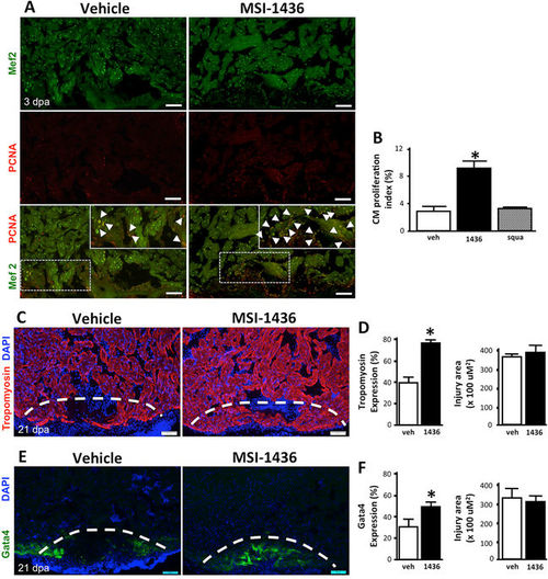
MSI-1436 stimulates adult zebrafish heart regeneration. a Representative images of cardiomyocyte proliferation 3 days post-amputation (dpa) of the ventricular apex in vehicle-treated fish and MSI-1436-treated fish. Arrows show proliferating cardiomyocytes expressing Mef2 and PCNA. b Quantification of proliferating cardiomyocytes 3 dpa expressed as percentage of cells Mef2-positive cells and PCNA-positive cells relative to cells expressing Mef2 only. Values are means ± S.E. (n = 10–12). *P < 0.01 compared to vehicle-treated fish. c Representative images of Tropomyosin expression in regenerating heart tissue 21 dpa. d Quantification of Tropomyosin expression and injury area 21 dpa. Values are means ± S.E. (n = 8–10). *P < 0.01 compared to vehicle-treated fish. e Representative images of Tg(gata4:GFP) expression in regenerating hearts at 21 dpa. f Quantification of Tg(gata4:GFP) expression and the injury area at 21 dpa. Values are means ± S.E. (n = 4-6). *P < 0.05 compared to vehicle-treated fish. Fish were given daily given intraperitoneal (IP) injections of either vehicle, 0.125 mg/kg MSI-1436 or 0.125 mg/kg squalamine (squa) following ventricular resection. Dashed lines show approximate resection plane |
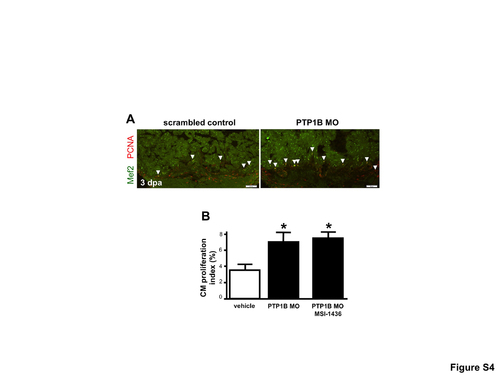
Antisense oligonucleotide mediated depletion of PTP1B enhances cardiomyocyte proliferation. (A) Representative images of cardiomyocyte proliferation 3 days post-amputation (dpa) of the ventricular apex in adult zebrafish treated with a scrambled control morpholino (MO) or a MO directed against PTP1B. Arrows show proliferating cardiomyocytes expressing Mef2 and PCNA. (B) Quantification of proliferating cardiomyocytes 3 dpa expressed as percentage of Mef2+PCNA+ cells relative to cells expressing Mef2 only. Values are means ± S.E. (n=9). *P<0.01 compared to control fish. Adult zebrafish were treated with daily intraperitoneal microinjections of scrambled control or PTP1B MO (1mg/kg) or PTP1B MO with MSI-1436. |
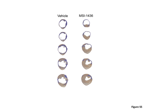
Effects of MSI-1436 on scar tissue formation. Representative heart sections from vehicle and MSI-1436 (0.125mg/kg) treated hearts were stained with acid fuchsin orange G staining to detect collagen (blue) and muscle (brown). |