
|
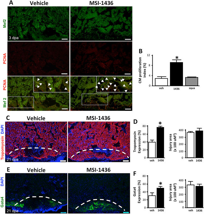
Fig. 2
MSI-1436 stimulates adult zebrafish heart regeneration. a Representative images of cardiomyocyte proliferation 3 days post-amputation (dpa) of the ventricular apex in vehicle-treated fish and MSI-1436-treated fish. Arrows show proliferating cardiomyocytes expressing Mef2 and PCNA. b Quantification of proliferating cardiomyocytes 3 dpa expressed as percentage of cells Mef2-positive cells and PCNA-positive cells relative to cells expressing Mef2 only. Values are means ± S.E. (n = 10–12). *P < 0.01 compared to vehicle-treated fish. c Representative images of Tropomyosin expression in regenerating heart tissue 21 dpa. d Quantification of Tropomyosin expression and injury area 21 dpa. Values are means ± S.E. (n = 8–10). *P < 0.01 compared to vehicle-treated fish. e Representative images of Tg(gata4:GFP) expression in regenerating hearts at 21 dpa. f Quantification of Tg(gata4:GFP) expression and the injury area at 21 dpa. Values are means ± S.E. (n = 4-6). *P < 0.05 compared to vehicle-treated fish. Fish were given daily given intraperitoneal (IP) injections of either vehicle, 0.125 mg/kg MSI-1436 or 0.125 mg/kg squalamine (squa) following ventricular resection. Dashed lines show approximate resection plane