FIGURE SUMMARY
Title

Zebrafish Infection: From Pathogenesis to Cell Biology

Authors
Torraca, V., Mostowy, S.
Source
Full text @ Trends Cell Biol.

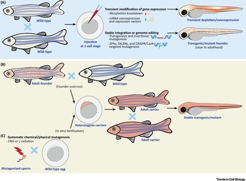
Approaches for Genetic Manipulation of Zebrafish. (A) Injection of constructs and chemicals in zebrafish eggs. Transient depletion can be performed by injection of morpholino oligonucleotides, RNA-binding oligomers that block translation/maturation of a specific (pre)-mRNA. Morpholinos can sometimes elicit off-target effects, therefore, it is important to validate phenotypes using alternative strategies and/or rescue experiments before conclusions can be fully drawn. Transient expression of genes can be obtained by injection of synthesized mRNA or plasmid DNA bearing an expression construct. Injected mRNAs will be expressed ubiquitously, while injection of plasmids enables cell- or tissue-specific expression. Zebrafish eggs can stably integrate DNA, which can be used to obtain stable transgenic lines or insertional mutants. The frequency of transgenesis is low when injecting DNA alone, but can be increased using transposases (i.e., Tol2) or meganucleases (i.e., I-SceI meganuclease). Zebrafish stable mutants can be efficiently generated with ZFNs, TALENs, or CRISPR/Cas9. These systems are based on induction of a site-specific double-stranded break, which is repaired via an error-prone non-homologous end joining mechanism. The CRISPR/Cas9 system has recently become the most common method to generate zebrafish mutants. Additionally, the CRISPR/Cas9 system has also been adapted to generate conditional/tissue-specific knockouts. Mutants are obtained by injecting mRNA or protein for the nuclease (together with guide RNA in the case of CRISPR/Cas9) in zebrafish eggs. Conditional/tissue-specific mutants are obtained by integration of a construct where Cas9 expression is controlled by an inducible or tissue-specific promoter. DNA constructs for stable integration can be designed with flanking homology recombination arms, which drive integration into a precise locus, and allow generation of knock-in lines. Generation of precise knock-in zebrafish is still challenging but can be facilitated by introducing double strand breaks at the site of interest (i.e., using TALENs or CRISPR/Cas9) [6]. Embryos manipulated using these techniques can be used for downstream functional studies, or, in the case of stable modifications, be raised to adulthood to establish a novel line. (B) Selection of stable transgenic or mutant lines. Carriers from (A) can be outcrossed to obtain heterozygote carriers. These offspring can be used for experiments, or raised to adulthood and inbred (i.e., to obtain homozygotes). (C) Systematic mutagenesis. Large libraries of random mutations can be obtained by exposure of sperm to chemical or physical mutagens (i.e., ENU or γ-radiation) prior to in vitro fertilization. Abbreviations: CRISPR/Cas9, clustered regulatory interspaced short palindromic repeats/CRISPR associated protein 9; ENU, N-ethyl N-nitrosurea; TALEN, transcription activator-like effector nuclease; ZFN, zinc-finger nuclease.

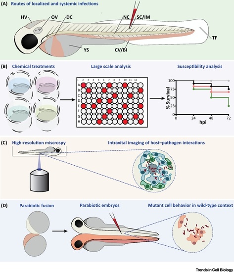
Methods for Studying Host–Pathogen Interactions Using Zebrafish. (A) Routes of zebrafish injection. Larvae can be injected locally into the YS or in body cavities, such as the HV and OV. Other compartments for injection include SC, IM, or the NC. HV, OV, IM, and TF infection all permit study of immune cell recruitment. The NC is inaccessible to immune cells but is valuable to model bone and cartilage inflammation. Injection into the circulation can be achieved by intravenous injections, for example via the CV/BI or the DC. This results in a rapid systemic dissemination of microbes throughout the body. (B) Chemical treatments. Zebrafish are suitable for toxicology research and for screening of libraries of bioactive compounds, including antimicrobials, because molecules in the bath water can be absorbed via the zebrafish skin. Survival and bacterial burden can be quantified to compare susceptibility of different genetic conditions or to assess the effect of chemicals/therapeutics in disease prevention. (C) Intravital imaging. Host–pathogen interactions can be followed
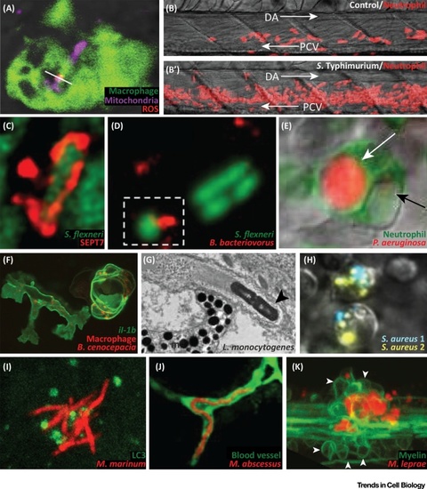
 in vivo by combining fluorescently-labeled bacteria and zebrafish transgenic lines reporting the expression pattern of specific genes or labeling specific cell types. A variety of proteins and subcellular compartments can also be tagged by fusing specific markers with fluorescent tags. (D) Parabiosis. Two zebrafish embryos can be fused by surgically forcing their blastulae into direct contact. This results in the development of conjoined embryos sharing blood circulation and body parts, enabling secreted factors and circulating cells to distribute in the bodies of both individuals. When applied to embryos with different genetic/transgenic makeup, this technique is useful to distinguish cell-autonomous from non-cell-autonomous functions. Abbreviations: BI, blood island; CV, caudal vein; DC, duct of Cuvier; HV, hindbrain ventricle; IM, intramuscular; NC, notochord; OV, otic vesicle; SC, subcutaneous; TF, tail fin; YS, yolk sac.

(A) Upon Salmonella Typhimurium infection, mitochondria (Mitotracker staining) produce ROS (MitoSOX staining) within macrophages (labeled with the macrophage-expressed gene 1 reporter Tg(Mpeg1:eGFP)gl22). White line: overlap between Mitotracker and MitoSOX. Adapted from [16]. (B, B′) Emergency granulopoiesis response in control (PBS injected) versus S. Typhimurium infected zebrafish larvae. Neutrophils are labeled with the lysozyme c reporter Tg(Lyz:DsRed)nz50. Arrows: direction of blood flow. Adapted from [20]. (C) Septin cage entrapment of Shigella flexneri visualized by SEPT7 immunolabeling. Adapted from [22]. (D) Bdellovibrio bacteriovorus predation of S. flexneri in the zebrafish hindbrain ventricle (outlined by broken box). Adapted from [28]. (E) Neutrophil labeled with the myeloperoxidase reporter Tg(Mpx:eGFP)uwm1) digesting Pseudomonas aeruginosa. White arrow: vacuole containing strong mCherry signal from P. aeruginosa. Black arrow: vacuole containing faint mCherry signal from P. aeruginosa. Adapted from [29]. (F) Macrophages (labeled with Tg(Mpeg1:mcherry-F)ump2) responding to Burkholderia cenocepacia infection with upregulation of interleukin-1β (labeled with the il-1b reporter Tg(il-1b:eGFP-F)zf550). Adapted from [33]. (G) Electron micrograph of Listeria monocytogenes (black arrowhead) propelled by an actin tail. Adapted from [35]. (H) Phagocytes infected with Staphylococcus aureus labeled with two different colors (S. aureus 1, S. aureus 2). Clearance of the inoculum will select a few persisters, leading to clonal selection. Adapted from [40]. (I) Recruitment of the autophagy marker LC3 (Tg(CMV:eGFP-LC3)zf155) to Mycobacterium marinum. Adapted from [60]. (J) Mycobacterium abscessus (R morphotype) presenting extracellular cording in a blood vessel (labeled with the kinase insert domain reporter Tg(kdr:eGFP)s843). Adapted from [66]. (K) Mycobacterium leprae infection, exacerbating neuronal damage, by altering axonal myelin (white arrowheads: myelin, labeled with the myelin basic protein reporter Tg(mbp:eGFP-CAAX)ue2). Adapted from [69]. Abbreviations: DA, dorsal aorta; m, melanophore; PCV, posterior cardinal vein; ROS, reactive oxygen species.

Acknowledgments
This image is the copyrighted work of the attributed author or publisher, and ZFIN has permission only to display this image to its users. Additional permissions should be obtained from the applicable author or publisher of the image. Full text @ Trends Cell Biol.