
- Title
-
Twa1/Gid8 is a β-catenin nuclear retention factor in Wnt signaling and colorectal tumorigenesis
- Authors
- Lu, Y., Xie, S., Zhang, W., Zhang, C., Gao, C., Sun, Q., Cai, Y., Xu, Z., Xiao, M., Xu, Y., Huang, X., Wu, X., Liu, W., Wang, F., Kang, Y., Zhou, T.
- Source
- Full text @ Cell Res.
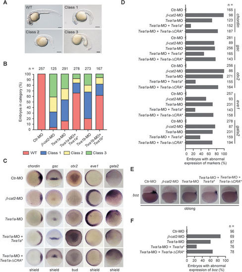
Zebrafish Twa1 is essential for dorsoventral patterning during embryogenesis. Zebrafish embryos at the one-cell stage injected with the indicated morpholinos targeting Twa1a and Twa1b (Twa1a-MO and Twa1b-MO) or mRNAs were harvested at the different times, and subjected to the following analyses. (A, B) Microscopy showed the different types of embryos at 24 h post-fertilization (hpf). The classes 1-3 represent increasing degrees of phenotypic severity in eye and brain structures. The percentages of embryos with the different classes are shown. (C-F) Whole-mount in situ hybridization of the dorsal (gsc, chordin and otx2) and ventral (gata2 and eve1) markers and the Wnt target gene boz. The percentages of embryos with abnormal expression of the indicated markers are also presented. β-Cat2, β-catenin 2; boz, bozozok; n, number of observed embryos; Twa1a*, zebrafish Twa1a-MO-resistant mRNA; Twa1a-ΔCRA*, zebrafish Twa1a-MO-resistant mRNA without CRA domain; Twa1b*, zebrafish Twa1b-MO-resistant mRNA. |

Characterization of zebrafish homologues of Twa1, Twa1a and Twa1b. (A) The deduced amino acid sequences of human Twa1 (hTwa1) and zebrafish Twa1a (Twa1a) and Twa1b (Twa1b). The CRA domain is underlined. (B) RT-PCR was performed on total RNAs isolated from the indicated developmental stages of zebrafish embryos. (C) The embryos were fixed at the indicated stages and processed for whole mount in situ hybridization with Twa1a and Twa1b probes. The embryos are shown in lateral view. hpf, hours post-fertilization; Prim-5, Primordia-5; Long-pec, Long-pectoral fin. |
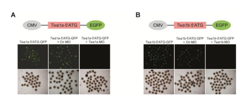
Effectiveness of the morpholinos targeting Twa1a and Twa1b mRNA (Twa1a-MO and Twa1b-MO) in zebrafish embryos. (A, B) The indicated MOs and constructs were injected into embryos at the one-cell stage. GFP signals were observed at 24 hpf. The plasmids contain zebrafish Twa1a- or Twa1b-5’ATG region carrying the targeting sequence of their corresponding MOs, which are fused with EGFP and driven by a cytomegalovirus (CMV) promoter. |