
- Title
-
Probing microtubules polarity in mitotic spindles in situ using Interferometric Second Harmonic Generation Microscopy
- Authors
- Bancelin, S., Couture, C.A., Pinsard, M., Rivard, M., Drapeau, P., Légaré, F.
- Source
- Full text @ Sci. Rep.
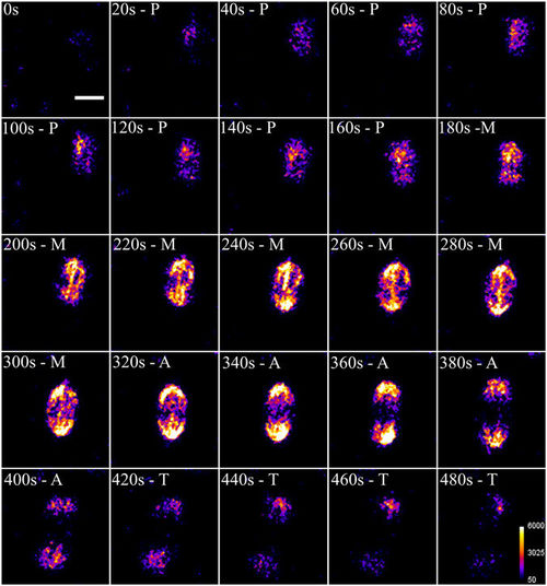
Time-lapse SHG imaging of mitotic spindles during mitotic division of a zebrafish embryo. Prometaphase (P; 20–160 s), metaphase (M; 180–300 s), anaphase (320–400 s) and telophase (420–480 s). Average power at focus: 150 mW, photomultiplier tube set at 1100 V. Scale bar: 10 µm. |
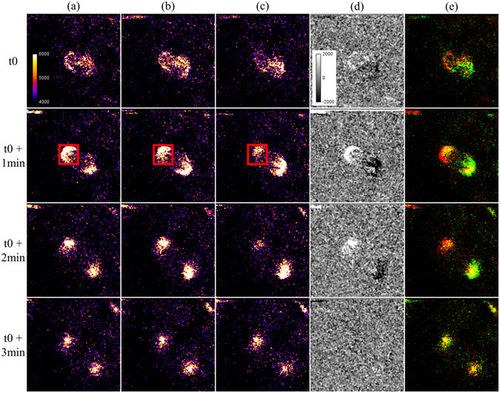
I-SHG of mitotic spindles in a zebrafish embryo. (a–c) Images acquired with different reference phases (0°, 90° and 180° respectively). Constructive and destructive interference occurs in opposite poles at 0° and 180° while no variations are observable at 90°. (d) Subtraction of the images (a,c) showing the interferometric contrast in the two poles. (e) 2-colors merge of the images acquired at 0° (in red) and 180° (in green). Average power at focus: 100 mW, photomultiplier tube set at 1050 V. Field of view: 50 × 50 µm2. |
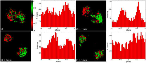
Image and histogram of the phase of the SHG signal generated in the spindles during mitosis. Field of view: 29 × 29 µm2. |