
- Title
-
Dietary lipid content reorganizes gut microbiota and probiotic L. rhamnosus attenuates obesity and enhances catabolic hormonal milieu in zebrafish
- Authors
- Falcinelli, S., Rodiles, A., Hatef, A., Picchietti, S., Cossignani, L., Merrifield, D.L., Unniappan, S., Carnevali, O.
- Source
- Full text @ Sci. Rep.

ZFIN is incorporating published figure images and captions as part of an ongoing project. Figures from some publications have not yet been curated, or are not available for display because of copyright restrictions. |
|
ZFIN is incorporating published figure images and captions as part of an ongoing project. Figures from some publications have not yet been curated, or are not available for display because of copyright restrictions. |

NUCB2/nesfatin-1-like ir in the gut of zebrafish fed with HDF-P and HFD-C compare to MFD-C. Cross sections of the zebrafish for nesfatin-1-like show higher abundance of nesfatin-1-like cell in HFD-C compared to HFD-P (3 F). For details on methods employed for percentage of positive intestinal epithelial cells calculation, please consult the Materials and Methods section. Values with different superscript letters are significantly different (p < 0.05). |
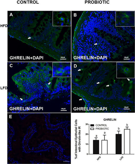
Ghrelin-like ir in the gut of zebrafish fed with HDF-P and HFD-C compare to LFD-C. Ghrelin positive cells did not show changes in the intestine of fish fed a LFD-C (4C) compare to LFD-P (4D) and HFD-C (4A) compare to HFD-P (4B). For details on methods employed for percentage of positive intestinal epithelial cells calculation, please consult the Materials and Methods section. Values with different superscript letters are significantly different (p < 0.05). EXPRESSION / LABELING:
|
|
ZFIN is incorporating published figure images and captions as part of an ongoing project. Figures from some publications have not yet been curated, or are not available for display because of copyright restrictions. |
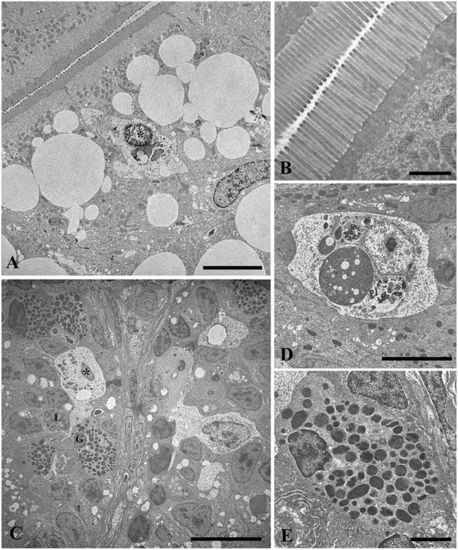
Elevated fat content increases lipid droplets, but did not alter intestinal mucosal epithelium in zebrafish fed a HFD, MFD and LFD. TEM micrograph showing large lipid droplets accumulation in the apical epithelium of enterocytes and intraepithelial macrophages in HFD-C fed fish (A). Intact brush border in HFD-C fed fish, (B). Macrophages in MFD-C fed fish in contact with the cell membrane of infiltrated lymphocytes and granulocytes (C). Intraepithelial macrophages with typical features of phagocytic cells in LFD-C fed fish (D). Magnification of intra-epithelium granulocytes in MFD-C fed fish (E). L = lymphocyte; G = granulocyte; Asterisk = macrophage. Scale bars: A = 5 µm, B = 1 µm, C = 10 µm, D = 5 µm, E = 2 µm. PHENOTYPE:
|

Lactobacillus rhamnosus did not alter intestinal mucosal epithelium in zebrafish fed a HFD-P, MFD-P and LFD-P. Micrographs showed the absence of damage signs in the intestine of fish fed with diet probiotic enriched (Figs. 6 A-F). Micrograph also showed an intact intestinal mucosal epithelium with intracellular tight junctions epithelial integrity, well organized microvilli and no cell debris in the intestinal lumen of zebrafish fed a HFD-P, MFD-P and LFD-P (Fig. 6 B, D, F). Scale bar: A, C, D ed E = 10 μm, B ed F = 1 μm |
|
Unillustrated author statements |