
- Title
-
Multicolor two-photon imaging of endogenous fluorophores in living tissues by wavelength mixing
- Authors
- Stringari, C., Abdeladim, L., Malkinson, G., Mahou, P., Solinas, X., Lamarre, I., Brizion, S., Galey, J.B., Supatto, W., Legouis, R., Pena, A.M., Beaurepaire, E.
- Source
- Full text @ Sci. Rep.
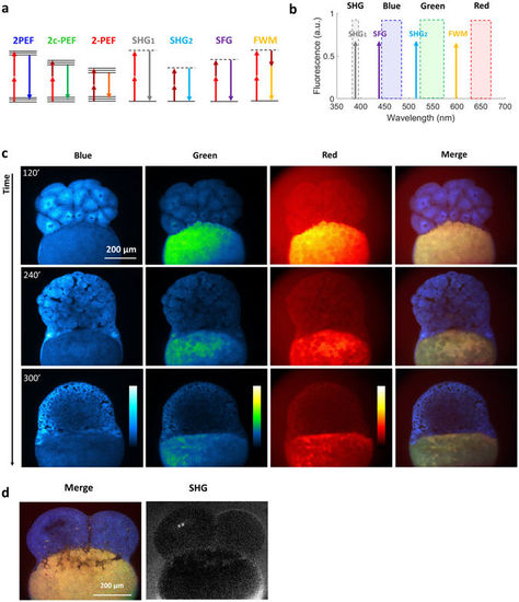
Multicolor two-photon efficient imaging of multiple endogenous fluorophores during early stages of zebrafish embryo development (a) Energy diagrams of two-photon excited fluorescence (2PEF) for blue and red fluorophores, two-color two-photon excited fluorescence (2c-2PEF) for green fluorophores, second harmonic generation (SHG), sum frequency generation (SFG) and four wave mixing (FWM). (b) Bandpass filters are chosen to select the emission of blue, green and red fluorophores rejecting coherent signals such as SHG, SFG and FWM. (c) Images of the blue, green, red fluorescence channel and SHG of the same zebrafish embryo at three different time points of development. Images of merged channels represent a shift in the spectroscopic characteristics of the yolk. (d) Multicolor two-photon and SHG images of the zebrafish embryo at 4 cell stage. Time per pixel, 40 µs. This experiment has been repeated in three independent samples. |
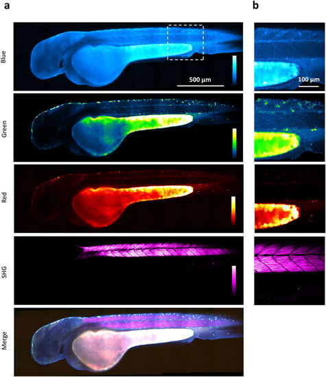
Multicolor two-photon efficient imaging of endogenous fluorophores in zebrafish embryo Images of the blue, green, red fluorescence, second harmonic generation, and merged channels of a live zebrafish embryo recorded 48 hours post fertilization (a) Large-scale (stitched mosaic) image encompassing 2450 × 730 µm2. (b) Detail extracted from the large-scale image. Pixel size, 0.83 µm. Multicolor pixel acquisition time, 40 µs. Scale bar, 500 µm. |
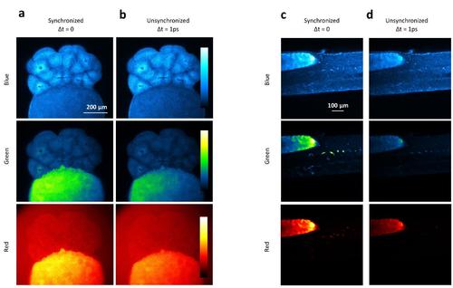
Enhancement of the green fluorescence by wavelength mixing in zebrafish embryo. Images of the blue, green, red, SHG and merged channels in zebrafish embryos at early stages of development (a-b) and at 48 hours developmental stage (c-d). Fluorescence images using synchronized (Δt=0) (b-d) and unsynchronized (Δt=1ps) pulse trains (a-c). |
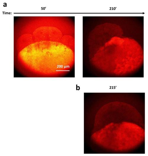
Decrease of the red autofluorescence is not due by photo-bleaching Red fluorescence channel of two embryos imaged from 8 stage cell (a) and from high state (b) |