FIGURE SUMMARY
Title

Utilizing Zebrafish Visual Behaviors in Drug Screening for Retinal Degeneration

Authors
Ganzen, L., Venkatraman, P., Pang, C.P., Leung, Y.F., Zhang, M.
Source
Full text @ Int. J. Mol. Sci.

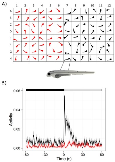
A typical visual motor response (VMR) experiment. Sufficient zebrafish embryos for an experiment are collected by breeding adult fish. The embryos can be maintained in petri dishes with media, as they develop until they are needed for a VMR assay. (A) At the appropriate stage, embryos can be placed into 96-well plate format to facilitate throughput, storage and, data collection during a VMR assay. It should be noted that there are multiple arrangements possible for placing zebrafish larvae in a 96-well plate, such as row-wise, column-wise, or checkerboard patterns. Larvae in the 96-well plate arrangement can then be placed in a light-proof recording chamber and exposed to light onset or light offset stimulus. The locomotor output of the larvae is recorded and processed. Recorded data can be visualized through programs such as R 3.4.0 [123] (B) This graph illustrates the VMR of a group of 7-dpf wild-type larvae (black trace) responding to light onset stimulus [106]. Their response is compared to the VMR from a group of visually-impaired pde6c mutant larvae (red trace). Healthy larvae exhibit a strong startle response to the light onset, while the visually-impaired larvae do not. This lack of response by retinal degeneration (RD) zebrafish models forms the basis for drug screens.

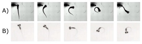
Zebrafish larvae display different startle escape behaviors. (A) A larva escapes from a touch-stimulus by exhibiting a C-bend. In this response, larvae curve their bodies in a C-shape and swim quickly away from the location of the stimulus [128]; (B) Larvae orient into an O-bend in response to a dark flash. The larva curves its body approximately 180 degrees to swim in the opposite direction [125]. Reproduced with permissions from Burgess et al. and Lorent et al.

Acknowledgments
This image is the copyrighted work of the attributed author or publisher, and ZFIN has permission only to display this image to its users. Additional permissions should be obtained from the applicable author or publisher of the image. Full text @ Int. J. Mol. Sci.