
- Title
-
Rare disruptive mutations in ciliary function genes contribute to testicular cancer susceptibility
- Authors
- Litchfield, K., Levy, M., Dudakia, D., Proszek, P., Shipley, C., Basten, S., Rapley, E., Bishop, D.T., Reid, A., Huddart, R., Broderick, P., Castro, D.G., O'Connor, S., Giles, R.H., Houlston, R.S., Turnbull, C.
- Source
- Full text @ Nat. Commun.
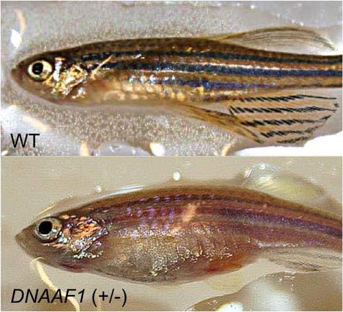
Comparison of wild-type male zebrafish and mutant DNAAF1Hu255h(+/-) zebrafish. On top is the healthy wild-type zebrafish compared to mutant DNAAF1Hu255h(+/-) zebrafish with visible tumor mass on bottom. TGCT was confirmed through histological characterization. PHENOTYPE:
|
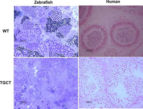
Histological characterisation of zebrafish and human tissue. On top left is age-matched (24–44 months old) wild-type zebrafish and on bottom left is the zebrafish DNAAF1Hu255h(+/-) tumor tissue, morphological tissue analysis of toluidine blue stained sections shows a dramatic loss of differentiated germ cells. On the top right for comparison is a healthy human testis and bottom right is a human seminoma. PHENOTYPE:
|
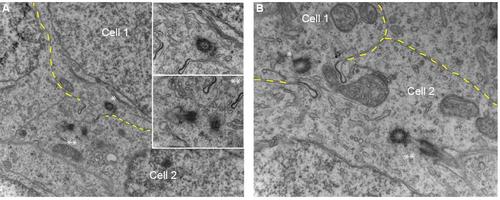
Ciliary structures adjacent to germ cell syncytium intercellular bridges from high resolution images of wild-type zebrafish male gonads. Spermatogonial stem cells commit to the development of spermatozoa via sequential steps of proliferation and differentiation after initial asymmetric cell division. A hallmark feature of spermatogonial derived early germ cell is the presence of intercellular bridges, concomitant from prematurely blocked cytokinesis followed by intercellular bridge stabilization. Presented are two gonadal sections displaying two adjacent male germ cell associated through intercellular bridges. Intriguingly, we observed presence of ciliary structures in the immediate vicinity of intercellular bridges. As the sections are ultrathin, only one of either cilium is visible as the corresponding cilium or centrosome is not in the same plane. A: The lower cell 2 shows mother and daughter centrioles where microtubules emanating from the mother centriole extend into a structure reminiscent of the primary vesicle (insert**). In cell 1, one centriole can be observed in the same plane B: Cell 2 shows presence of a cilium clearly featured by an axonemal structure composed of a membrane wrapped microtubule structure. Cell membranes are indicated in yellow. |