
Image
|
Figure Caption
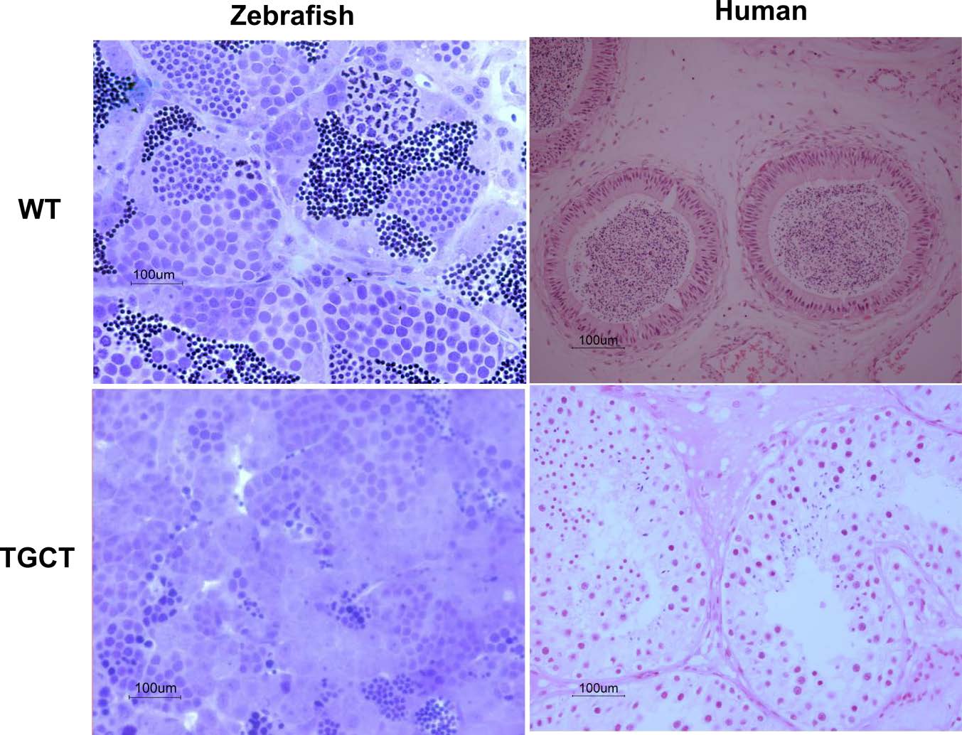
Fig. S2b
Histological characterisation of zebrafish and human tissue. On top left is age-matched (24–44 months old) wild-type zebrafish and on bottom left is the zebrafish DNAAF1Hu255h(+/-) tumor tissue, morphological tissue analysis of toluidine blue stained sections shows a dramatic loss of differentiated germ cells. On the top right for comparison is a healthy human testis and bottom right is a human seminoma.
Figure Data
Acknowledgments
This image is the copyrighted work of the attributed author or publisher, and
ZFIN has permission only to display this image to its users.
Additional permissions should be obtained from the applicable author or publisher of the image.
Full text @ Nat. Commun.