
- Title
-
FOXE3 contributes to Peters anomaly through transcriptional regulation of an autophagy-associated protein termed DNAJB1
- Authors
- Khan, S.Y., Vasanth, S., Kabir, F., Gottsch, J.D., Khan, A.O., Chaerkady, R., Lee, M.W., Leitch, C.C., Ma, Z., Laux, J., Villasmil, R., Khan, S.N., Riazuddin, S., Akram, J., Cole, R.N., Talbot, C.C., Pourmand, N., Zaghloul, N.A., Hejtmancik, J.F., Riazuddin, S.A.
- Source
- Full text @ Nat. Commun.
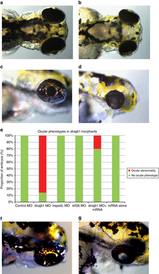
In vivo modelling reveals broader developmental defects and underdeveloped cataractous lenses in dnajb1a morphants. dnajb1a zebrafish morphants recapitulate PA symptoms. Dorsal view of (a) control morphant and (b) dnajb1a morphant embryos suggest a relatively compact lens observed at 4 days post fertilization as a result of dnajb1a MO. Lateral view of (c) control morphant and (d) dnajb1a morphant embryos showing broader eye developmental defects including underdeveloped cataractous lenses in dnajb1a MO-injected embryos. (e) Quantification of the compact lens phenotype in the dnajb1a, hspa4l and kif3a morphants and rescue of morphant phenotype with zebrafish orthologue of dnajb1a mRNA. Note: kif3a MO did not show any ocular phenotype; however, the morphants exhibited morphological and developmental defects, such as body curvature, consistent with the ciliary phenotypes. Lateral view of (f) control morphant and (g) dnajb1b morphant embryos illustrate a relatively compact cataractous lenses observed at 4 days post fertilization as a result of dnajb1b translation-blocking MO. PHENOTYPE:
|
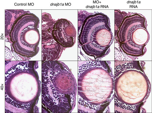
Histological evaluation of the developmental defects in the anterior segment of dnajb1a morphant eyes. Zebrafish embryos injected with the dnajb1a sbMO (5 ng), or the morpholino plus dnajb1a mRNA (150 pg) or dnajb1a mRNA. Embryos were fixed 4 days post fertilization in 4% paraformaldehyde and embedded in paraffin. Sections (5 µm) were stained with haematoxylin and eosin and visualized under a microscope for histological evaluation. PHENOTYPE:
|

dnajb1 morphants display significant apoptosis in the eye. TUNEL staining of control embryo sections (a-f) and dnajb1 morphants (g-l) injected with the dnajb1a sbMO (5 ng). The dnajb1a morphants show significantly increased apoptotic nuclei throughout the eye compared with the control embryo, which was completely devoid of TUNEL-positive nuclei. The morphant lens extends out of the retina and is fused to the cornea with an increased number of nuclei as compared with the control (white arrows, h,k) and is characterized by the presence of apoptotic nuclei in the centre of the lens (white arrows, i,l), suggesting an abnormal lens development. Note: the control embryo section image was acquired at a higher exposure time compared with the morphant to produce a background. PHENOTYPE:
|
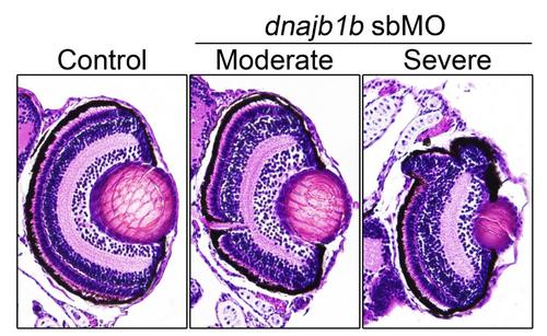
Histological evaluation of the developmental defects in the anterior segment of dnajb1b morphant eyes. Zebrafish embryos were injected with a morpholino (10ng), and 4-days post fertilization, embryos were fixed in 4% paraformaldehyde and embedded in paraffin. 5µm sections were stained with hematoxylin and eosin. The less severe morphant embryos showed protruding lenses while the severely affected morphant embryos exhibited smaller eyes with somewhat elongated/ bulged lenses compared to the controls. PHENOTYPE:
|

Unillustrated author statements PHENOTYPE:
|