
- Title
-
Natural Bizbenzoquinoline Derivatives Protect Zebrafish Lateral Line Sensory Hair Cells from Aminoglycoside Toxicity
- Authors
- Kruger, M., Boney, R., Ordoobadi, A.J., Sommers, T.F., Trapani, J.G., Coffin, A.B.
- Source
- Full text @ Front. Cell. Neurosci.
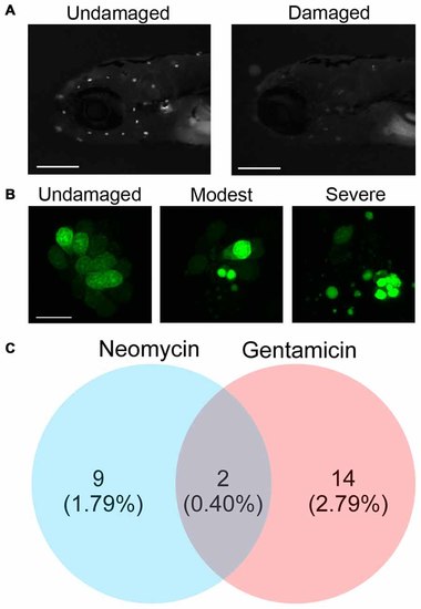
Screening a natural compound library for otoprotective compounds. (A) Examples of larval zebrafish labeled with the vital dye Yo-Pro-1. The left image is of an untreated fish, while the right image shows reduced neuromast fluorescence caused by exposure to 200 μM neomycin. Scale bars = 250 μm. (B) High magnification images of the SO2 neuromast from an untreated fish (left), fish with moderate hair cell damage (center), or fish with severe hair cell damage (right). Damage is evident by nuclear condensation, which results in more intense, punctate labeling, and by fragmentation. The scale bar on the left image = 10 μm and applies to all three panels. (C) Natural compounds protect zebrafish lateral line hair cells from the aminoglycosides neomycin and gentamicin. The initial screen produced nine compounds that protected hair cells from neomycin, 14 from gentamicin, and two from both ototoxins. The majority of the natural compounds screened (477) were not protective. |
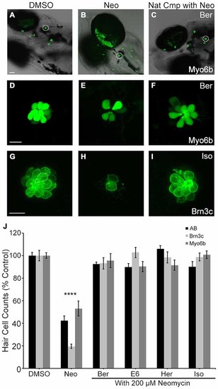
Direct hair cell counts confirm otoprotection. Ber, E6, Her and Iso robustly protect hair cells from 200 μM neomycin after acute exposure. (A–F) Representative low magnification (A–C) and high magnification (D–F) images of myo6b: EGFP transgenic fish treated with the indicated conditions. White circles in (A–C) denote the neuromasts shown in (D–F). Scale bar in (A) = 40 μm and applies to panels (A–C), scale bar in (D) = 10 μm and applies to panels (D–F). (G–I) Representative images of brn3c: mGFP hair cells treated with the indicated conditions. (G) The dotted line encircles a hair cell. Scale bar in (G) = 10 μm and applies to panels (G–I). (J) Direct counts of hair cells in five neuromasts per fish demonstrate significant hair cell protection in *AB fish labeled with anti-parvalbumin (black bars, one-way ANOVA, F (4,60) = 46.58, p < 0.0001), Brn3c fish (light gray bars, one-way ANOVA, F(4,38) = 75.58, p < 0.0001), and myo6: EGFP fish (dark gray bars, one-way ANOVA, F(4,53) = 14.86, p < 0.0001). N = 8–24 and error bars are ±SEM. and ****p < 0.0001. |
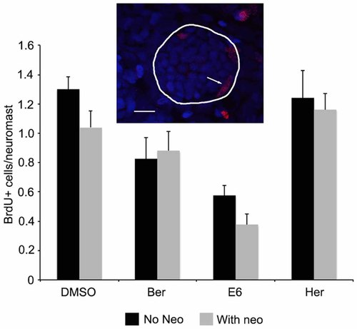
Ber, E6, and Her do not enhance regeneration rates after neomycin damage. Fish were treated with either natural compound only or natural compound and 200 μM neomycin in the presence of BrdU. Fish were counterstained with DAPI to identify neuromasts based on characteristic morphology, as shown in the inset where DAPI-labeled nuclei are blue and red nuclei are BrdU+. The line encircles the neuromast and the arrow denotes a BrdU+ supporting cell at the neuromast periphery. Scale bar = 10 μm. There is no difference in the number of BrdU+ cells per neuromast in fish that received neomycin + compound (gray bars) vs. those that received compound only (black bars; 2-way ANOVA, treatment effect F(1,67) = 1.882, p = 0.175). N = 8–10 and bars are ±SEM. |
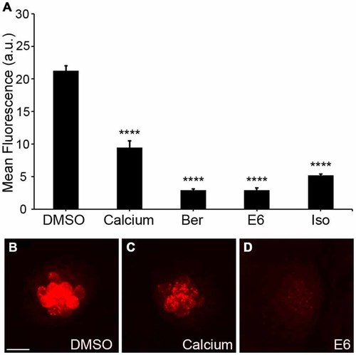
Natural compounds reduce uptake of gentamicin conjugated with Texas Red (GTTR). (A) The OPC of Ber, E6, or Isosignificantly reduced fluorescent intensity of 50 μM GTTR after 18 min of GTTR incubation (one-way ANOVA, F(4, 69) = 179.0, p < 0.0001). Fluorescent intensity was measured in arbitrary units (a.u.). (B–D) Representative images of 18 min GTTR uptake in DMSO (negative control), high Ca2+ (positive control), and E6. Scale bar in (B) = 10 μm and applies to all images. N = 13–15 and bars are +SEM. and ****p < 0.0001. |
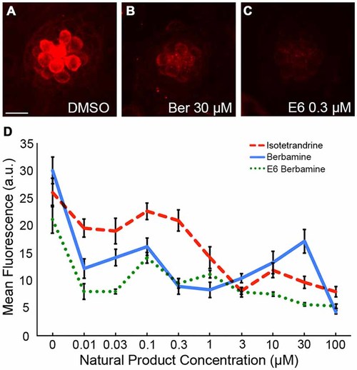
Natural compounds reduce FM 1-43FX entry into hair cells. (A–C) Representative images showing a reduction in FM 1-43FX uptake with Ber and E6. The scale bar in (A) = 10 μM and applies to all images. (D) Ber, E6, and Iso reduce uptake of FM 1-43FX into hair cells in a dose-dependent manner. Fluorescent intensity was measured in arbitrary units (a.u.). Data were analyzed by one-way ANOVA for each compound, with p < 0.0001 in all cases. N = 10–14 and bars are ±SEM. |