
- Title
-
Id4 functions downstream of Bmp signaling to restrict TCF function in endocardial cells during atrioventricular valve development
- Authors
- Ahuja, S., Dogra, D., Stainier, D.Y., Reischauer, S.
- Source
- Full text @ Dev. Biol.
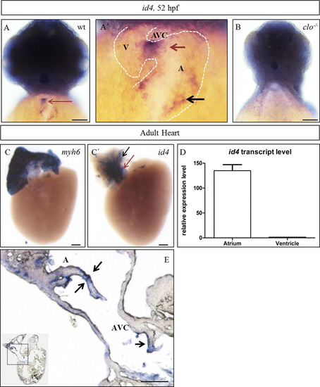
id4 expression analysis in zebrafish embryos and adult hearts. (A; A′) id4 is expressed in the brain and heart at 52 hpf. In the heart, expression is enriched in the AVC (red arrow) and inflow tract (black arrow). (B) Lack of id4 expression in clo mutant hearts suggests that id4 is expressed in the endocardium. (C; C′) In the adult heart, id4 is expressed in the atrium (black arrow) and AV canal (red arrow) exclusively. myh6 (atrial mhc) was used as a positive control for the atrium. (D) qPCR analysis of id4 expression in the adult zebrafish heart. (E) Section through adult heart after whole mount in situ hybridization. Arrows point to endocardial expression of id4 in the adult atrium and AVC. Scale bars, 100 µm. A, atrium; AVC, atrioventricular canal; V, ventricle. |
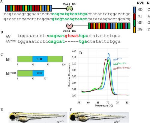
Generation of an id4 loss-of-function allele. (A) Left and right TALEN arms attached with FokI DD and FokI RR were targeted to the genomic region corresponding to the bHLH domain of Id4. (B) id4bns18, a 5 bp deletion allele, was recovered post TALEN mutagenesis. (C) This 5 bp deletion is predicted to lead to the formation of a 72-amino-acid-long truncated protein (p.Val73*). (D) Alterations in the High Resolution Melt analysis were used to determine the id4 genotype. (E, F) Overall morphology of id4-/- larvae is indistinguishable from their id4+/+ siblings at 72 hpf. RVD: repeat-variable diresidue, N: Nucleotide. |
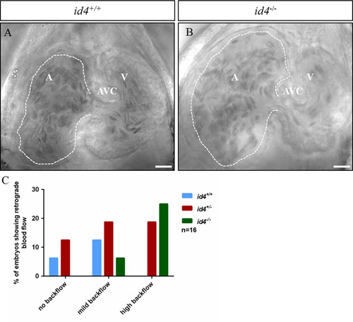
id4 mutant larvae exhibit retrograde blood flow after heat stress conditions. Embryos from an incross of id4 heterozygous animals were exposed to heat stress at 48 hpf for 1 h at 37 ºC, followed by bright field spinning disk microscopy. (A, B) Single frames (at end of ventricular systole) from blood flow movies of id4+/+ and id4-/- siblings respectively. (C) Quantification of embryos showing retrograde flow. Scale bars, 20 µm. A, Atrium; AVC, Atrioventricular Canal; V, Ventricle. Blood flow movies of id4+/+ (id4 wt video 1) and id4-/- (id4 mut video 2) are available in supplementary information. PHENOTYPE:
|
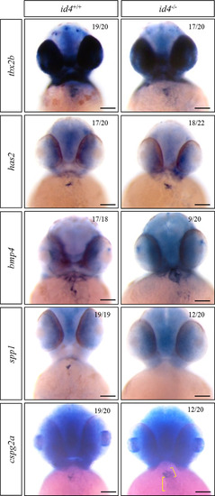
AV canal marker gene expression analysis in id4 mutants at 52 hpf. Myocardial (tbx2b, bmp4, cspg2a) and endocardial (has2, spp1) AVC marker gene expression was analyzed in embryos from incrosses of id4 heterozygous animals. After in situ hybridization, embryos were separated according to expression patterns and subsequently genotyped. Amongst these markers, tbx2b and has2 expression were found to be unchanged, bmp4 and cspg2a were misexpressed and spp1 expression was reduced in id4 mutants. Scale bars, 100 µm. |
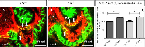
Expression of Alcam in AV endocardial cells is expanded in id4 mutants. Alcam positive AV endocardial cells (yellow) were found to be increased in id4 mutants (B) as compared to their id4 heterozygous siblings (A) at 52 hpf. (C) Quantification of Alcam positive AV endocardial cells. (A, Atrium; AVC, Atrioventricular Canal; V, Ventricle; SAV, Superior AVC; IAV, Inferior AVC). Scale bars, 20 µm. p-value **- 0.0016, *- 0.0114. (Green – Alcam; Red - Tg(kdrl:ras-mCherry). |
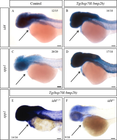
Id4 mediates Bmp signaling leading to spp1 expression. Increased expression of id4 (A, B) and spp1 (C, D) was observed at 52 hpf in Tg(hsp70l:bmp2b) embryos after heat shock at 26 and 48 hpf for 30 min at 37 ºC. (E, F) spp1 expression was examined in id4 wild-type siblings and mutants in the Tg(hsp70l:bmp2b) background at 52 hpf after previously mentioned heat shock conditions. Unlike their id4+/+ siblings (E), most of the id4-/- (F) did not show an increase in spp1 expression after heat shock induced bmp2b overexpression. Scale bars, 100 µm. |
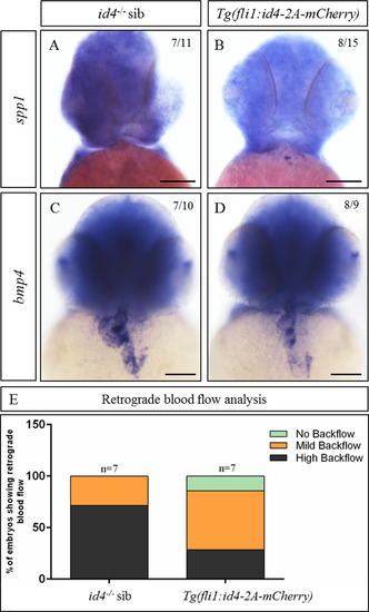
Endothelial overexpression of id4 in id4 mutants can partially rescue their valve phenotype. Although id4-/- (A) generally lack spp1 expression, a subset does exhibit spp1 expression after endothelial specific id4 overexpression (B). Similarly, as compared to id4-/- which show ectopic bmp4 expression (C), id4 mutants with endothelial specific id4 overexpression (D) show reduced misexpression of bmp4. (E) Most of the larvae with endocardial re-expression of id4 in the id4-/- background show mild retrograde blood flow as compared to id4-/- siblings, most of which show high backflow. Scale bars, 100 µm. |
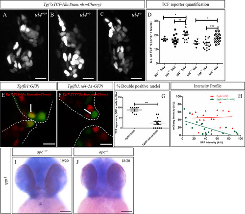
Id4 negatively regulates TCF function in the atrioventricular canal (AVC). (A, B, C) Larvae from incrosses of id4 heterozygous fish or from crossing id4+/- males to id4-/- females in a Tg(7xTCF-Xla.Siam:nlsmCherry) background were imaged at 77 hpf followed by quantification (D) of TCF positive nuclei. We observed a significant increase in the number of TCF reporter positive cells in id4 mutants as compared to siblings. (E, F) Transient endothelial specific overexpression of id4 caused a dose dependent reduction of TCF reporter signal (F) in the AVC as compared to the overexpression of GFP alone (E). Arrows point to GFP expressing TCF reporter positive nuclei (E) and id4 expressing TCF reporter negative nuclei (F). (G) Percentage of TCF reporter positive cells in transient GFP and id4 overexpression larvae and their intensity profile (H). (I, J) At 52 hpf, apc mutants show reduced spp1 expression as compared to their wild-type siblings. Scale bars, A-C and E-F: 10 µm; I-J: 100 µm (SAV, Superior AVC; IAV, Inferior AVC.) p value *<0.05, **<0.01, ***<0.001. EXPRESSION / LABELING:
PHENOTYPE:
|
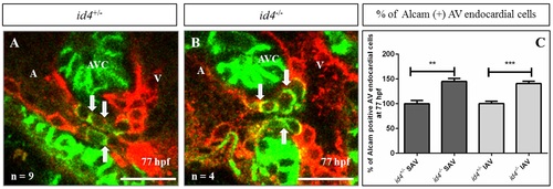
Expression of Alcam in AV endocardial cells is expanded in id4 mutants. Alcam positive AV endocardial cells (indicated by arrows) were found to be increased in id4 mutants (B) as compared to their id4 heterozygous siblings (A) at 77 hpf . (C) Quantification of Alcam positive AV endocardial cells. (A, Atrium; AVC, Atrio-Ventricular Canal; V, Ventricle; SAV, Superior AVC; IAV, Inferior AVC) Scale bars, 20 µm. p-value **- 0.0016, ***- 0.0003. (Green - Alcam; Red - Tg(kdrl:ras-mCherry). |
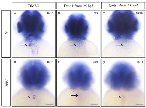
Bmp inhibitor Dmh1 treatment leads to reduction of id4 and spp1 expression. Wild-type embryos were treated with 10 µm Dmh1 from 25 or 35 hpf until 52 hpf. Decreased expression of id4 (B,C) and spp1 (E,F) was observed in Dmh1 treated embryos as compared to DMSO treated ones where expression pattern of id4 (A) and spp1 (D) appeared wild-type like. Scale bars, 100 µm. |
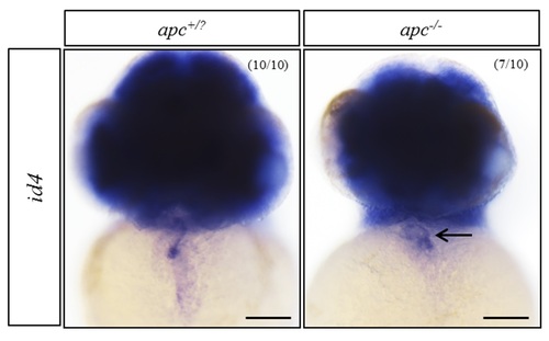
id4 expression is expanded into the ventricle inapcmutants at 52 hpf. id4 expression was observed in the ventricle at 52 hpf in apc mutants, unlike in the wild-type siblings which showed id4 expression restricted to the AVC. Scale bars, 100 µm. |
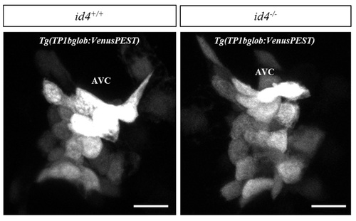
Notch signaling appears to be unaffected in id4 mutants at the AVC. Representative maximum intensity projections of id4+/+ and id4-/- AVC in Notch signaling reporter background. No significant difference was observed. Scale bars, 10 µm. |
Reprinted from Developmental Biology, 412(1), Ahuja, S., Dogra, D., Stainier, D.Y., Reischauer, S., Id4 functions downstream of Bmp signaling to restrict TCF function in endocardial cells during atrioventricular valve development, 71-82, Copyright (2016) with permission from Elsevier. Full text @ Dev. Biol.