
- Title
-
Graded effects of unregulated smooth muscle myosin on intestinal architecture, intestinal motility, and vascular function in zebrafish
- Authors
- Abrams, J., Einhorn, Z., Seiler, C., Zong, A.B., Sweeney, H.L., Pack, M.
- Source
- Full text @ Dis. Model. Mech.
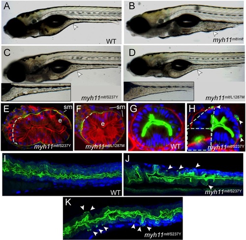
Modifier mutations induce cell invasion in mlt heterozygotes. (A-D) Lateral views of live 6-dpf larvae. Arrowhead points to the mid-intestine. (A) A wild-type (WT) larva. (B) A homozygous mlt larva in which there is invasive expansion of the mid- and posterior intestine. (C) A mlt/S237Y compound heterozygote. Arrowhead points to a region of altered intestinal morphology. Inset shows a more severely affected mlt/S237Y compound heterozygote. (D) mlt/L1287M compound heterozygote. Mild and moderate (inset) phenotypes are shown as in C. (E,F) Histological section through the posterior intestine of 6-dpf mlt/S237Y and mlt/L1287M compound heterozygotes following their immunostaining with anti-Keratin (red) and anti-Laminin-1 antibodies (green). Dashed line indicates predicted position of basement membrane disrupted by the invasive epithelial cells; e, intestinal epithelium; sm, smooth muscle. (G,H) Histological section through the posterior intestine of 3-dpf Tg(miR194-Lifeact-GFP) wild-type and sibling mlt/S237Y larvae following immunostaining with anti-GFP (green) and anti-Laminin-1 (red) antibodies, showing GFP-labeled invadopodia (arrowheads) of epithelial cells extending through gaps in the basement membrane. Inset shows a higher-magnification view. (I-K) Confocal projections through the mid- and posterior intestine of 4-dpf Tg(miR194-Lifeact-GFP) wild-type and sibling mlt/S237Y (myh11mlt/S237Y) larvae following anti-GFP immunostaining (green). Arrowheads point to invadopodia protruding from the basal epithelial cell plasma membrane in the mlt/S237Y mutants. Blue, DAPI-stained nuclei. EXPRESSION / LABELING:
PHENOTYPE:
|
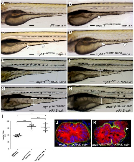
mlt modifier mutants are sensitized to oxidative stress. (A-D) Lateral views of 82-hpf wild-type (WT), homozygous mlt, heterozygous mlt and L1287 homozygous larvae. Wild-type (A), mlt heterozygote (C) and L1287M homozygote (D) larvae treated with menadione (1.5µM; mena). Morphological changes of the mid- and posterior intestine that are characteristic of epithelial invasion (arrowheads) are seen in the mlt larva (B) and the menadione-treated mlt heterozygote and L1287M homozygote (C,D). In all three mutant larvae, the ventral intestine appears to extend into the yolk. Intestinal morphology is normal in the wild-type larva (A). (E-H) Lateral views of 6-dpf KRAS-axin larvae that either have homozygous wild-type myh11 (E,F), S237Y (G) or L1287M (H) alleles. The intestine is expanded in KRAS-axin larvae as a result of epithelial hypertrophy (brackets, E,F). Further expansion as a result of epithelial invasion is seen in menadione-treated KRAS-axin S237Y and L1287M homozygotes (brackets, G,H), as previously reported for menadione-treated KRAS-axin mlt heterozygotes (Seiler et al., 2012). (I) Quantification of intestinal width in KRAS-axin compound-mutant larvae treated with menadione compared with untreated siblings. ****P<0.001; n.s., not significant. Unpaired Student′s t-test performed; mean±s.e.m. (J,K) Histologic analyses of immunostained larvae showing epithelial cell invasion in the intestine of a menadione-treated homozygous S237Y KRAS-axin larva (K) vs a wild-type KRAS-axin larva (J). Green, anti-Laminin-1; red, anti-Keratin; blue, DAPI-stained nuclei. Arrowhead (K) points to invasive cells. Scale bars: 100µm. EXPRESSION / LABELING:
PHENOTYPE:
|

Zebrafish myh11 mutations alter intestinal transit. (A,B) Intestinal transit as measured by the expulsion of fluorescent microspheres in wild-type and sibling homozygous mlt larvae injected with a myh11 morpholino (MO) (A), and wild-type and sibling S237Y homozygous larvae (B). Anterior: beads remain in anterior intestinal bulb; posterior: beads remain in mid- and posterior intestine; expelled: no beads remaining in the intestine. ****P<0.001. Chi-squared test performed with 2 degrees of freedom. (C) Time lapse of a wild-type (WT) larva beginning when the fluorescent beads are present in the anterior intestine and followed until bead expulsion. A, M, P, location of anterior intestine, mid-intestine and posterior intestine, respectively. PHENOTYPE:
|

ZFIN is incorporating published figure images and captions as part of an ongoing project. Figures from some publications have not yet been curated, or are not available for display because of copyright restrictions. PHENOTYPE:
|
|
Unillustrated author statements PHENOTYPE:
|