
- Title
-
BPIQ, a novel synthetic quinoline derivative, inhibits growth and induces mitochondrial apoptosis of lung cancer cells in vitro and in zebrafish xenograft model
- Authors
- Chiu, C.C., Chou, H.L., Chen, B.H., Chang, K.F., Tseng, C.H., Fong, Y., Fu, T.F., Chang, H.W., Wu, C.Y., Tsai, E.M., Lin, S.R., Chen, Y.L.
- Source
- Full text @ BMC Cancer

Effect of BPIQ on proliferation of NSCLC tumor cells. a The structures of CPT and BPIQ. b Three NSCLC H1299, A549 and H1437 cells were incubated with various concentrations of BPIQ for 24 and 48 h, respectively. The percentage of viable cells was calculated as a ratio of BPIQ- to DMSO-treated control cells. c The tumor volume in the zebrafish xenograft model. The intensity of red fluorescence is proportional to the xenograft tumor size. N = 20 embryos for each group. d The quantificative analysis of c. All data are presented as mean ± S.D. of three independent experiments. (*p < 0.05, **p < 0.005 and ***p < 0.001 against vehicle control, respectively) |
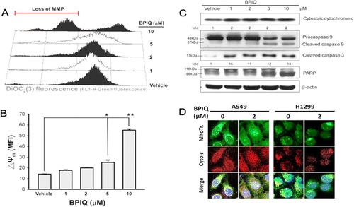
Loss of MMP and caspase activation by BPIQ. a H1299 cells were exposed to media containing the indicated concentrations of BPIQ or vehicle control for 24 h, stained with DiOC2(3), then analyzed for changes in their fluorescent profile by flow cytometry. b Quantitative analysis. Data are presented as means ± S.D. Histograms represent one of three independent experiments. *p < 0.05 and **p < 0.001 against vehicle control, respectively. c Western blot analysis demonstrating BPIQ-induced cytochrome c release and cleavage of caspase-9 and 3, as well as PARP. β-actin was measured as an internal control. d The distribution of cytochrome c in the cytosol of two NSCLC cell lines A549 and H1299 following 2 µM BPIQ treatment. ª mitochondria; ª cytochrome c; ª DAPI; ª co-localization of mitochondria and cytochrome c. Magnification 200 x |