
- Title
-
Nr2f1b control venous specification and angiogenic patterning during zebrafish vascular development
- Authors
- Li, R.F., Wu, T.Y., Mou, Y.Z., Wang, Y.S., Chen, C.L., Wu, C.Y.
- Source
- Full text @ J. Biomed. Sci.

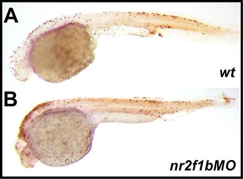
Expression of nr2f1b in during zebrafish development. a At 15S, nr2f1b is expressed in the telencephalon (t), ventral medial diencephalon (d), hindbrain rhombomeres (h) and lateral plate mesoderm (lpm, arrow) corresponding in location to the developing vasculature. At 20hpf (~24S), nr2f1b is expressed in the vessels (b). c, c′ At 24 hpf, nr2f1b is expressed in the telencephalon (t), diencephalon (d), hindbrain (h), as well as in vessels (v), and caudal vein plexus (CVP) of the trunk. c′ is an enlargement of C. d, e Cross sections of embryos from c′ show that nr2f1b is expressed in dorsal aorta (da), posterior cardinal vein (pcv), and caudal vein plexus (CVP). Scale bars in all figures represent 100 µm. EXPRESSION / LABELING:
|
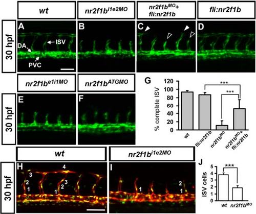
Morpholino knockdown of nr2f1b causes defects in vascular development. a In uninjected control embryos, the arota (da) and posterior cardinal vein (pcv) have formed by 30 hpf and intersegmental vessels (isv) have reached the DLAV at the dorsal aspect of the embryo. At the same stage ISVs are stalled mid-somite in nr2f1b i1e2 (b), nr2f1b e1i1 (e) and nr2f1b ATG (f) morphants. Overexpression of nr2f1b has no obvious defect in vasculature (d), but rescues the defect of ISV stalling (solid arrowhead) as shown in (c). g Quantification of percentage of completed ISV shows a 40 % increase compare to nr2f1b morphants (*** refers to p < 0.0001 by an unpaired student′s t-test. Scale bars are 50 µm for (a-f). h and i Imaging of endothelial nuclei in green and vessels in red at 30 hpf in wild-type control and nr2f1b MO-treated embryos using Tg(fli1a:nEGFP) y7 :(kdrl:mCherry) ci5 double transgenic line. nr2f1b morphants showed reduced ISV nuclei numbers (i). j Quantification of ISV nuclei number in nr2f1b morphants (n = 18) compared to wild-type control (n = 16). ***P < 0.001, Student t test. |
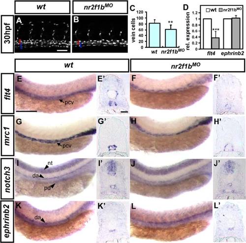
Nr2f1b modulates vein cell number and marker expression. a-c At 30 hpf, loss of nr2f1b function in Tg(fli1a:negfp) y7 embryos results in reduced vein cell number (b, blue bracket) as compared to uninjected wild type controls (a). The artery is marked by a red bracket. c Quantitative analysis shows a significant reduction in vein cell number in nr2f1b morphants. Compared to wild type controls. e, g, i, k, expression of the venous markers flt4 (f) and mrc1 (g) is reduced in the trunk of nr2f1b morphants at 24 hpf while there is no obvious change in the expression of arterial markers notch3 (i) and ephrinb2 (k). e′-l′ are cross sections of embryos in (e-l). d Quantification by qPCR shows a 60 % reduction in flt4 expression but no change in ephrinb2 expression in nr2f1b morphants (***refers to p < 0.0001 and **refers to p < 0.001 by an unpaired student′s t-test. Scale bars represent 50 µm in a, b, e-l and scare bar in cross-sections e′-l′ is 30 µm). Abbreviations: posterior cardinal vein (pcv), dorsal aorta (da), neural tube (nt) and pronephric ducts (pd). |
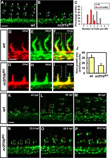
Nr2f1b is required for the growth of intersegmental vessels. The number of cells forming each ISV were counted in wild type control Tg(fli1a:negfp) y7 (a) and nr2f1b morphant embryos (b) at 30 hpf. c Proportional distribution of ISVs containing 1-7 cells in both conditions. d-j Time-lapse imaging of wild type Tg(kdrl:eGFP) la116 (d-f) and nr2f1b morphant (g-i) embryos to examine the extension of ISV tip cell filopodia. Confocal images at 23 hpf (red; d, g) and 26hpf (green; e, h) were merged (f, i). The extension of tip cell was quantitated by pixel intensity and shows reduced extension of tip cell in nr2f1bMO (j). k-p Time-lapse imaging of wild type Tg(fli1a:nEGFP) y7 (k-m) and nr2f1b morphant (n-p). Scale bars represent 50 µm. PHENOTYPE:
|
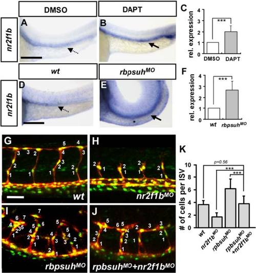
The expression of Nr2f1b is upregulated by notch signaling. a, b nr2f1b expression is increased at 24 hpf embryos after treatment with DAPT as compared to DMSO control embryos by in situ hybridization. d, e nr2f1b expression is upregulated in rbpsuh morphants at 24 hpf embryos. c, f Quantification by qPCR showed the increased expression of nr2f1b in rbpsuh morphants or DAPT treated embryos at 24 hpf significantly. g-j Representative confocal images showing the number of nuclei per ISV at 30 hpf in g wild-type (wt) embryos, h nr2f1b MO, i rbpsuh MO, and j rbpsuh MO with nr2f1b MO. k Quantitation of the average number of cells per ISV in single and double morphants. ***refers to p < 0.0005 by an unpaired student′s t-test. Scale bars represent 100 µm in a, b, d, e and 50 µm in g-j. |
An increase in non-specific cell death after morpholino injection is not the cause of the observed vascular phenotype TUNEL labeling was used to detect apoptotic cells in wild type and nr2f1b morphants. Some cell death was observed at the head region, but cell death in vascular regions was not elevated over that observed in wild type controls. |
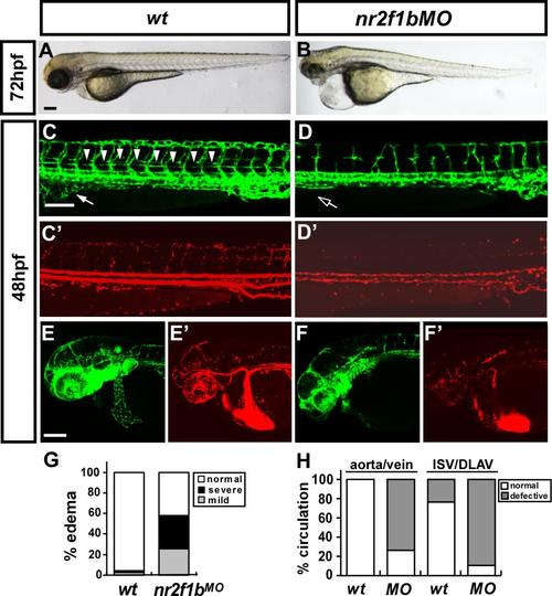
Loss of nr2f1b results in pericardial edema, absent parachordal vessels, subintestinal vessels (SIV) mispattern and circulation defects. (A,B) nr2f1bMO injected embryos showed severe pericardial edema at 72hpf compared to wt, in addition, there is an obvious smaller eyes in nr2f1b morphants. Quantitatively, 60% of nr2f1b morphants (n=43) with mild to severe pericardial edema compared to wt (n=49) as shown in (G). (C-F, C′-F′) nr2f1bi1e2 MO was injected into transgenic Tg(fli:eGFPy1;gata1:dsRedsd2) embryos with GFP-labeled endothelial cells (C,D,E,F) and dsRed-labeled blood cells(C′,D′,E′,F′). nr2f1b morphants show mispatterned ISV, DLAV and SIV (arrow), and absent parachordal vessels (arrowhead) (C and D) and circulation defect at the ISV and DLAV and slow to lose axial circulation of the aorta and vein (D′) in the trunk region at 48hpf as compared to wild-type (C′). Vasculature and circulation defects are also observed in the head region of nr2f1b morphants (F and F′) as compared to wt (E and E′). Quantification of the number of embryos exhibiting trunk vessel circulation defects in wt (n=8) and nr2f1b morphants (n=25) as shown in (H). The scale bars in C-F and C′-F′ represent 100 µm and in A-B are 500 µm. PHENOTYPE:
|