FIGURE SUMMARY
Title

Cross-species models of human melanoma

Authors
van der Weyden, L., Patton, E.E., Wood, G.A., Foote, A., Brenn, T., Arends, M.J., Adams, D.J.
Source
Full text @ J. Pathol.

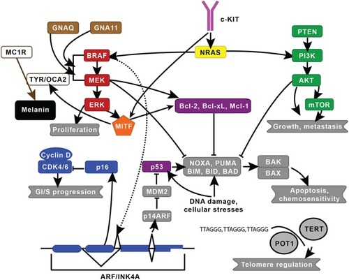
Established melanoma pathways. The two major signalling pathways implicated in melanoma are the mitogen‐activated protein kinase and the phosphatidylinositol‐4,5‐bisphosphate 3‐kinase pathways, which are in red and green, respectively. Key genes include c‐KIT (pink), CDK (blue), GNAQ/GNA11 (brown), MITF (orange), NRAS (yellow), and P53/BCL (purple). MC1R, which is involved in skin pigmentation, and TERT and POT1, which are involved in telomere regulation, are also shown. This figure was modified from Vidwans et al142 under the Creative Commons Attribution License. The lines shown indicate known interactions between pathways or molecules.

Naevi and melanomas driven by oncogenic forms of Braf and NRAS. (A, B) Naevi developing in adult BrafV618E mice 48. Naevi generally became visible 6–8 weeks after the induction of BrafV618E expression. (C, D) Melanoma from the same BrafV618E model. D shows an invasive malignant melanoma with evidence of infiltration and destruction of the overlying surface epithelium and invasion into the subcutaneous adipose tissue. The average latency to melanoma formation was 426 days in this model. (E) H&E‐stained section of an ocular melanoma, with melanoma cell infiltration of the lens and the subretinal tissues, that developed in a 13‐week‐old Tyr::NRASQ61K mouse 135. Original magnification × 50.

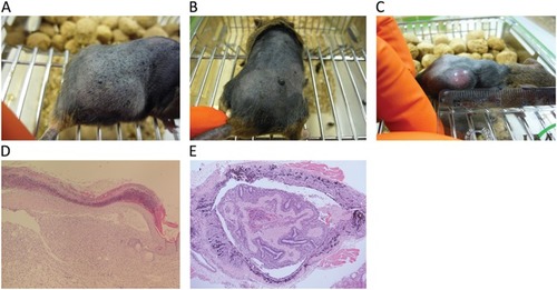
Canine, equine, and zebrafish melanoma. (A) A canine melanoma developing in the nasal cavity and (B) spreading to the viscera, particularly the liver. (C) An equine melanoma showing multinodular dermal lesions around the tail base and masses expanding into the pelvic canal and regional nodes. (D) An equine spleen with multiple malignant melanomas, and liver and lymph node from the same case. (E) Melanomas arising in a BRAFV600E; mitf zebrafish and (F) in a BRAFV600E; p53 zebrafish. The photographs in A and B were kindly provided by Jeff Caswell, Department of Pathobiology, University of Guelph, Guelph, Ontario, Canada N1G2W1.

Acknowledgments
This image is the copyrighted work of the attributed author or publisher, and ZFIN has permission only to display this image to its users. Additional permissions should be obtained from the applicable author or publisher of the image. Full text @ J. Pathol.