
- Title
-
INSL3 stimulates spermatogonial differentiation in testis of adult zebrafish (Danio rerio)
- Authors
- Assis, L.H., Crespo, D., Morais, R.D., França, L.R., Bogerd, J., Schulz, R.W.
- Source
- Full text @ Cell Tissue Res.
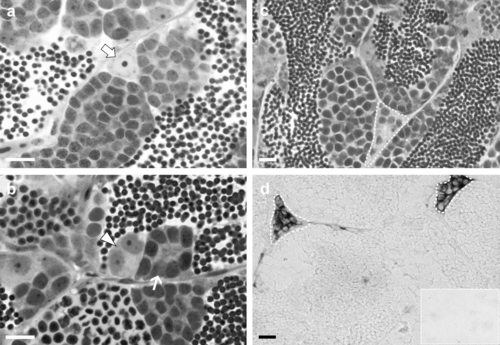
Morphological characteristics of zebrafish type A undifferentiated (Aund) and type A differentiating (Adiff) and type B spermatogonia and Leydig cells and in situ hybridization for insl3 mRNA in adult zebrafish testis. a Type Aund spermatogonia (arrow) are single germ cells showing a large and clear nucleus, with poorly condensed chromatin and one or two compact nucleoli. b A cyst containing two type Adiff spermatogonia (arrowhead) that show smaller nuclei stained more intensely by toluidine-blue. Type B spermatogonia (thin arrow) have still smaller, slightly elongated/ovoid nuclei that moreover show a high amount of heterochromatin. c Interstitial space (dashed line) with a group of Leydig cells. d Adult zebrafish testis section showing the detection of insl3 mRNA by in situ hybridization in Leydig cells cluster (dashed lines). Inset in d non-specific staining obtained with the sense probe. a-c Sections were prepared for morphological analysis according to Leal et al. (2009b). Magnification ×1000 (a, b), ×600 (c, d). Bars 10 µm EXPRESSION / LABELING:
|
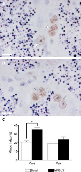
a, b Testis tissue sections showing BrdU-positive germ cells: type Aund (arrow), type Adiff (thin arrows) and type B (arrowhead) spermatogonia. Magnification ×1000. Bars 10 µm. c Mitotic indices of type Aund and Adiff spermatogonia after incubation for 7 days in the absence (Basal) and presence (hINSL3) of 100 ng hINSL3/ml. *Significant difference (P < 0.05) between treated and control. Results are presented as means ± SEM (n = 8) |
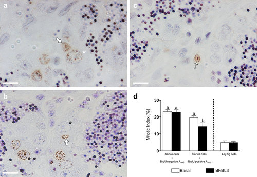
a, b Testis tissue sections showing BrdU-positive Sertoli cells nuclei (arrows) in association with BrdU-positive (white star) and BrdU-negative (black star) type Aund spermatogonia. c BrdU-positive Leydig cell nucleus (thin arrow). d Mitotic indices of Sertoli cells in association with BrdU-negative or BrdU-positive type Aund spermatogonia and of Leydig cells after incubation for 7 days in the absence (Basal) or presence (hINSL3) of 100 ng hINSL3/ml. Different letters indicate significant differences (P < 0.05) between the absence and presence of hINSL3. Magnification ×1000 (a-c). Bars 10 µm. Results are presented as means ± SEM (n = 8) |