
- Title
-
Effective heritable gene knockdown in zebrafish using synthetic microRNAs
- Authors
- Giacomotto, J., Rinkwitz, S., Becker, T.S.
- Source
- Full text @ Nat. Commun.
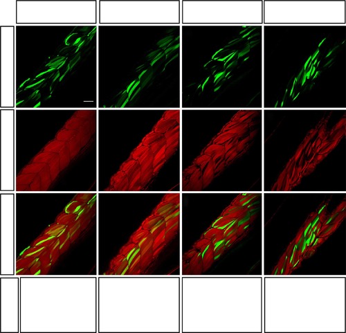
Validation of knockdown efficiency of three miRNA-expressing constructs in 52 hpf zebrafish embryos. (a–l) Homozygous βactin:mCherry:sUTR embryos were injected with 30pg of three different tol2-flanked miRNA-expressing constructs, along with transposase to promote integration. Each construct was designed to produce GFP along with the same specific artificial miRNA targeting a unique site in 3′-UTR of mCherry. Control animals were injected with the empty mmu-miR155 backbone. A minimum of 10 larvae per condition were analysed using a confocal microscope to estimate knockdown efficiency. (m–p) Homozygous βactin:mCherry:sUTR embryos were injected with corresponding RNA (150pg)—synthesized via linearized pME-RNAi clones—and raw red fluorescence was quantified, normalized and presented as percentage of control. The means of 20 embryos±s.e.m. Different from control at *Pd0.02, **P=0.0001, ***P=0.000001. Scale bar, 50µm. |
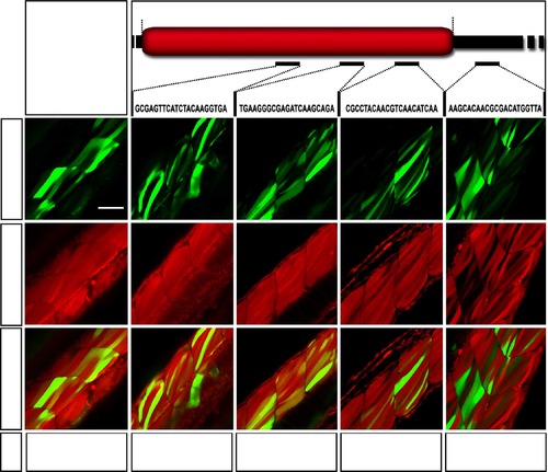
Evaluation of the effect of target sequence/position on knockdown efficiency in 52 hpf zebrafish embryos. (a) Schematic representation of mCherry mRNA produced in the βactin:mCherry:sUTR zebrafish line, as well as in artificial miRNA targets. (b–p) Embryos with ubiquitous mCherry expression were injected with transposase and 30pg of tol2-flanked miRNA-expressing constructs (mmu-miR155 backbone), which were designed to produce artificial miRNA targeting different sites of the mCherry mRNA. A minimum of 10 larvae per condition were analysed in order to estimate knockdown efficiency. (q–u) Homozygous βactin:mCherry:sUTR embryos were injected with corresponding RNA (150pg), synthesized via linearized pME-RNAi clones, and raw red fluorescence was quantified, normalized and presented as percentage. The means of 10 embryos±s.d. Different from control at *P=0.05, **P=0.000001. Scale bar, 50µm. |

ZFIN is incorporating published figure images and captions as part of an ongoing project. Figures from some publications have not yet been curated, or are not available for display because of copyright restrictions. EXPRESSION / LABELING:
PHENOTYPE:
|
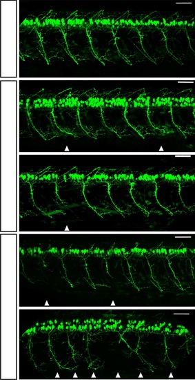
Representative images of 52 hpf zebrafish larvae expressing GFP in motor neurons ± smn1 knockdown. Lateral views of (a) control (empty vector) and (b-e) transgenic zebrafish larvae expressing artificial miR (targeting smn1 3′-UTR). Compared with control animals, transgenic larvae expressing miRsmn1 present abnormal motor neuron development, including abnormal branching, short axons as well as pathfinding errors (white arrowheads). Transgenic animals expressing artificial miRNA against the ORF (targets 2 and 3) do not show obvious motor neuron abnormality. Scale bar, 50µm. |
|
ZFIN is incorporating published figure images and captions as part of an ongoing project. Figures from some publications have not yet been curated, or are not available for display because of copyright restrictions. PHENOTYPE:
|
|
ZFIN is incorporating published figure images and captions as part of an ongoing project. Figures from some publications have not yet been curated, or are not available for display because of copyright restrictions. PHENOTYPE:
|
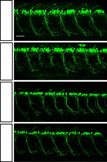
hsa-SMN1 rescues the dre-smn1 knockdown motor neuron phenotype. Representative images of 52hpf zebrafish larvae expressing GFP in motor neurons. Lateral views of (a) control (empty vector) transgenic animals. (b–d) transgenic UBI:miRsmn1-1#5 zebrafish line (presenting ~90% downregulation of SMN), with hsa-SMN1 RNA injection (c) or hsa-SMN1 ubiquitous expression via integrated transgene (d; see Supplementary Fig. 7 for strategy used). Scale bar, 50µm. EXPRESSION / LABELING:
PHENOTYPE:
|
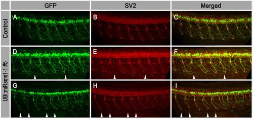
Representative images of 52hpf hsa-miR218-2:GFP zebrafish larvae after immunostaining against GFP and SV2 proteins. A-C, Lateral views of control (empty vector) larvae. D-I, UBI:miRsmn1-1#5 transgenic larvae expressing an artificial miRNA targeting the 3′UTR of smn1 transcripts. Motor neuron abnormalities, such as short axons, abnormal branching and/or pathfinding errors, are labeled by white arrowheads. |
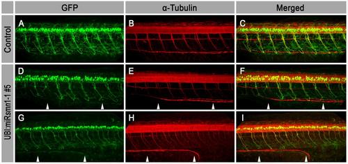
Representative images of 52hpf hsa-miR218-2:GFP zebrafish larvae after immunostaining against GFP and a-tubulin proteins. A-C, Lateral views of control (empty vector) larvae. D-I, UBI:miRsmn1-1#5 transgenic larvae expressing an artificial miRNA targeting the 3′UTR of smn1 transcripts. Motor neuron abnormalities, such as short axons, abnormal branching and/or pathfinding errors, are indicated by white arrowheads. |
|
ZFIN is incorporating published figure images and captions as part of an ongoing project. Figures from some publications have not yet been curated, or are not available for display because of copyright restrictions. PHENOTYPE:
|