
|
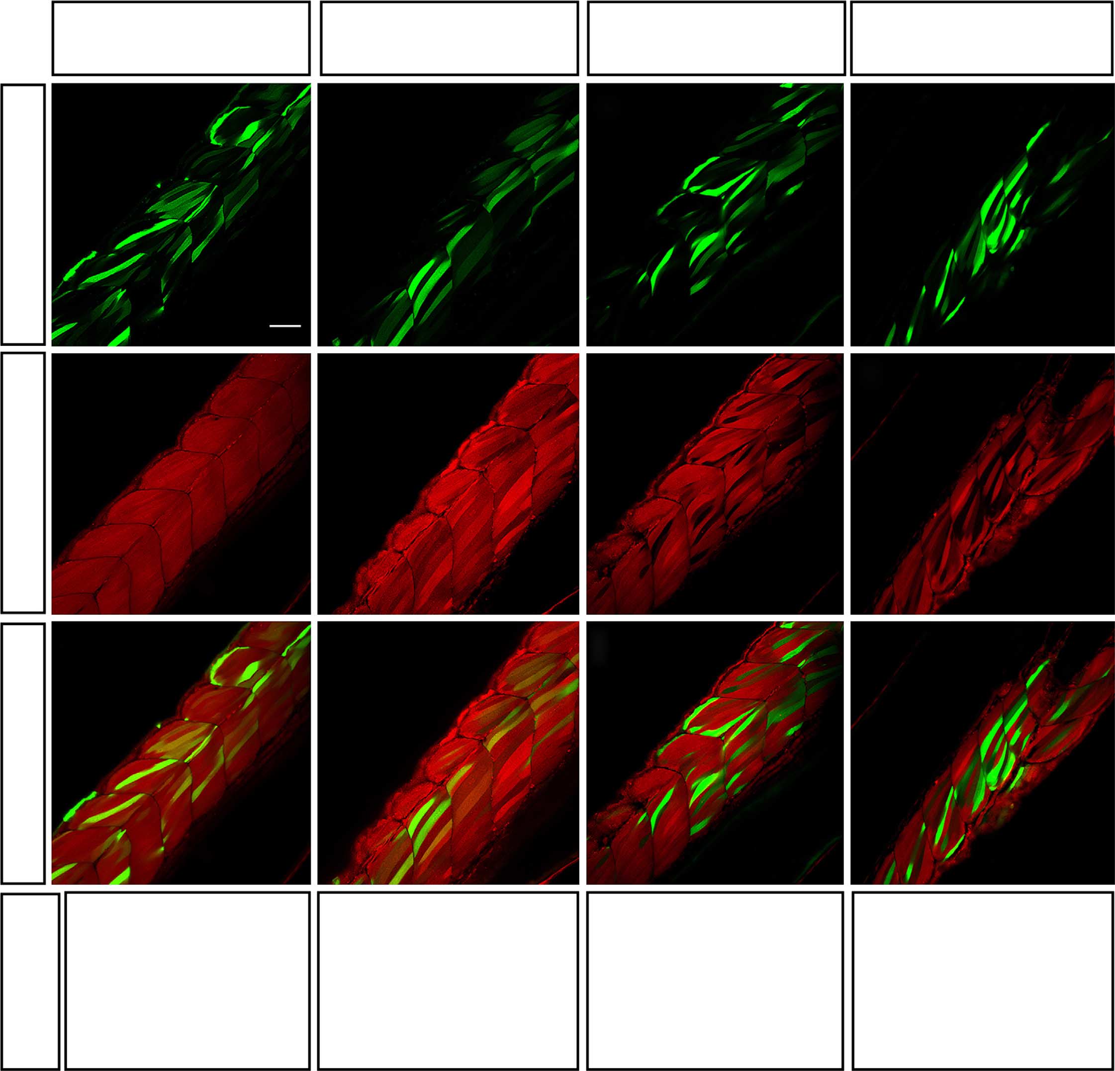
Fig. 2 Validation of knockdown efficiency of three miRNA-expressing constructs in 52 hpf zebrafish embryos.
(a–l) Homozygous βactin:mCherry:sUTR embryos were injected with 30pg of three different tol2-flanked miRNA-expressing constructs, along with transposase to promote integration. Each construct was designed to produce GFP along with the same specific artificial miRNA targeting a unique site in 3′-UTR of mCherry. Control animals were injected with the empty mmu-miR155 backbone. A minimum of 10 larvae per condition were analysed using a confocal microscope to estimate knockdown efficiency. (m–p) Homozygous βactin:mCherry:sUTR embryos were injected with corresponding RNA (150pg)—synthesized via linearized pME-RNAi clones—and raw red fluorescence was quantified, normalized and presented as percentage of control. The means of 20 embryos±s.e.m. Different from control at *Pd0.02, **P=0.0001, ***P=0.000001. Scale bar, 50µm.