
- Title
-
On the Crucial Cerebellar Wound Healing-Related Pathways and Their Cross-Talks after Traumatic Brain Injury in Danio rerio
- Authors
- Wu, C.C., Tsai, T.H., Chang, C., Lee, T.T., Lin, C., Cheng, I.H., Sun, M.C., Chuang, Y.J., Chen, B.S.
- Source
- Full text @ PLoS One
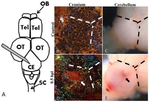
The diagram of the stab lesion assay. A: Schematic diagram of the stab lesion. A 27G syringe was used to create a cranial wound of depth 1.5 mm in the cerebellum (OB, olfactory bulb; Tel, telencephalon; OT, optic tectum; Ce, cerebellum; SC, spinal cord). B–E: Bright field images of the cranium (B, D) and exposed brains (C, E) of control fish and after the stab lesion. B and C show intact cranium and cerebellum of the control (no stab lesion) and D and E show the injured cranium and cerebellum of an experimental animal at 0.5 hour post-lesion (hpl). The fresh wound can be seen clearly on both the cranium and the cerebellum (D, E; the white arrow shows the lesion site on the cranium and the black arrow shows the wound on the exposed cerebellum). |
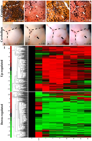
Regeneration of cerebellum at different day-post-lesion and the time-course microarray data in cerebellar wound healing process. A-H: Bright field images of the craniums (A, C, E, G) and cerebellums (B, D, F, H) at different time points post lesion. The cranial and cerebellum were intact before stab lesion (A, B). At 3dpl, the wound can be seen on the cranial and cerebellum. (C, D) At 7dpl, the cranial is seal and scar is observed in the cerebellum. (E, F) At 14 dpl, the scar was hardly seen. (Scale bar, 100 μm.). (I) The differentially expressed genes (≥1.5 or ≤0.67 fold change) are hierarchically clustered (5839 genes). Columns represent the day-post-lesion (dpl), and the rows represent the genes. The blocks indicate temporally up-regulated and down-regulated genes, respectively. The color bar represents the log value of the ratio relative to the intensity at 0 dpl. |
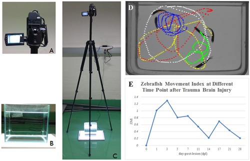
Equipment setup for behavior analysis. A: Close-up views of the video camera. B: Close-up views of the aquarium. C: The camera and aquarium setup. D: Swim sample paths of zebrafish after TBI. E: Quantitative behavior index (zebrafish movement index, ZMI) of injured zebrafish describes the degree of disability in the behavior of the injured zebrafish. The greater the index value, the greater the degree of disability. |