
- Title
-
Basal body proteins regulate Notch signaling via endosomal trafficking
- Authors
- Leitch, C.C., Lodh, S., Prieto-Echagüe, V., Badano, J.L., Zaghloul, N.A.
- Source
- Full text @ J. Cell Sci.
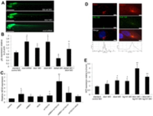
Loss of BBS proteins and genetic interaction with endosomal components enhances Notch pathway activation. (A) GFP fluorescence in representative Tp1blgob:eGFP transgenic embryos at 48hours post fertilization (hpf) in bbs1 morphant (MO) embryos and nicd-mRNA-injected embryos. Scale bar: 0.5mm (B) qRT-PCR quantification of GFP mRNA levels relative to β-actin in 24hpf Tp1blgob:eGFP embryos. *Pd0.01 compared with standard control morpholino; **Pd0.005 compared with deltaA morpholino (Student′s t-test). (C) qRT-PCR analysis of relative HES5 expression in HEK293 cells expressed as the fold change relative to control cells. Data represent the mean±s.d. (seven separate experiments). *Pd0.01 compared with control; **Pd0.01 compared with either short hairpin alone (Student′s t-test). (D) Double immunofluorescent labeling of either early endosomes (EEA1) or recycling endosomes (RAB11) and primary cilia (ARL13B or acetylated tubulin) in hTERT-RPE1 cells. Histograms represent the intensity of red fluorescence across the image. Scale bars: 5 μm. (E) qRT-PCR analysis showing relative GFP expression in Tp1bglob:eGFP zebrafish embryos at 24 hpf. Data show the mean±s.d. *Pd0.005 compared with standard control morpholino; **Pd0.005 compared with morpholinos alone (Student′s t-test). EXPRESSION / LABELING:
|

Notch1 localization in ciliated and unciliated cells and suppression of Notch-inhibited cell type markers in bbs4 morphants. (A) hTERT-RPE1 cells doubled immunostained for detection of NOTCH1 (green) and ARL13B (red) and transfected with or without shBBS4. 40X magnification. Cilia in ciliated cells (+) magnified in inset boxes. Scale bars = 20 μm. (B) Expression of neurogenin1 in control MO-, bbs4 MO- and tsg101 MO-injected embryos at 24 hpf detected by whole mount in situ hybridization. (C) Expression of shippo1 in control MO-, bbs4 MO- and tsg101 MO-injected embryos at 24 hpf. Insets indicate magnification of dashed boxes. EXPRESSION / LABELING:
|