
- Title
-
Zebrafish etv7 regulates red blood cell development through the cholesterol synthesis pathway
- Authors
- Quintana, A.M., Picchione, F., Klein Geltink, R.I., Taylor, M.R., and Grosveld, G.C.
- Source
- Full text @ Dis. Model. Mech.
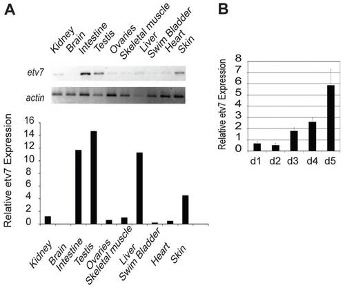
etv7 is expressed in the adult and developing zebrafish. (A) Semi-quantitative RT-PCR of etv7 mRNA of adult zebrafish tissues. actin is provided as a positive loading control. Quantification of the etv7 signals relative to actin is shown below. (B) Quantitative real-time PCR measuring the level of etv7 expression during the first 5 days of development. All values are relative to day 0, which is 4 hpf. d1=day 1 post-fertilization etc. Error bars represent standard deviation. |
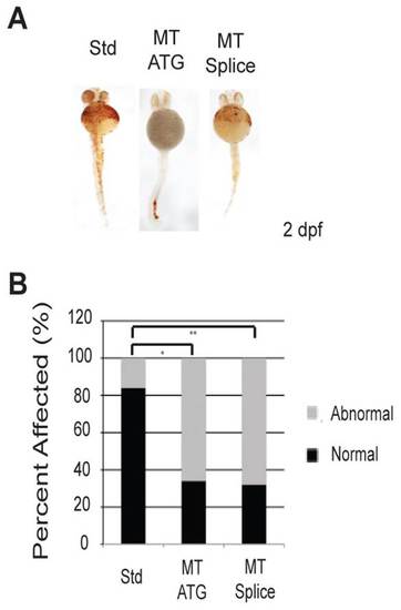
Loss of etv7 causes a marked reduction in hemoglobinized red blood cells. (A) etv7 morphants stained with o-dianisidine at 2 dpf. Std represents embryos injected with 8.2 ng of standard control morpholino, MT ATG are embryos injected with 8.2 ng of translation-blocking morpholino, and MT Splice are embryos injected with 4.1 ng of splice-site morpholino. Normal embryos were classified by the presence of adequate heme staining according to o-dianisidine, and fish with reduced heme staining are classified as abnormal. (B) Quantitation of A (MT ATG n=44, Std n=44, MT Splice n=34). *Pd0.0001, **Pd0.0001. PHENOTYPE:
|
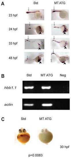
etv7 morphants express beta-globin (hbbe1.1). (A) In situ hybridization analyzing the expression of beta-globin mRNA was performed on embryos injected with 8.2 ng of standard control morpholino (Std) or 8.2 ng of translational-blocking morpholino (MT ATG) at 23, 24, 33 or 48 hpf. Arrows indicate expression in the posterior blood island (PBI) and arrowheads depict expression on the yolk sac. (B) Semi-quantitative PCR of beta-globin (hbbe1.1) expression at 24 hpf. actin expression is shown as loading control. Std are animals injected with 8.2 ng of standard control morpholino, MT ATG are animals injected with 8.2 ng of translational blocking morpholino and Neg is control PCR reaction without template. (C) etv7 morphants exhibit reduced levels of mature red blood cells. o-dianisidine staining was performed at 30 hpf with embryos injected with 8.2 ng of translational-blocking morpholino (MT ATG) or 8.2 ng standard control morpholinos (Std). EXPRESSION / LABELING:
PHENOTYPE:
|

gata1 expression is maintained at 48 hpf in morphants. In situ hybridization detecting gata1 mRNA expression at 23, 24, 33 and 48 hpf in standard control-injected embryos (Std) or etv7 translational-blocking morpholinos (MT ATG). 8.2 ng of each morpholino was injected. Arrows indicate expression in the posterior blood island (PBI) and arrowheads depict expression on the yolk sac. EXPRESSION / LABELING:
|
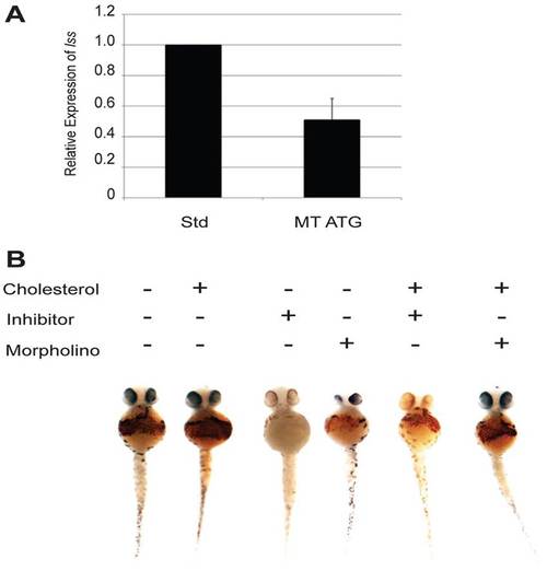
etv7 regulates red blood cells through the cholesterol synthesis pathway. (A) Real-time PCR analysis of lss expression in embryos injected with either 8.2 ng of standard control morpholino (Std) or 8.2 ng of etv7 morpholinos (MT ATG). Error bars represent standard deviation. (B) Inhibition of Lss enzymatic activity phenocopies knockdown of etv7. Wild-type embryos were treated with the Lss inhibitor Ro 48-8.071 (50 nM) and stained with o-dianisidine at 2 dpf. Injection of etv7 morpholinos was performed simultaneously and compared with embryos treated with Ro 48-8.071. Rescue experiments were performed by injecting cholesterol into the yolk of embryos at 1 dpf. o-dianisidine staining was used to visualize the presence or absence of hemoglobinized red blood cells at 2 dpf (Std n=26, Std with cholesterol n=32, MT ATG n=20, MT ATG with cholesterol n=15). Refer to Table 1 for P-values and percent affected in each category. PHENOTYPE:
|

In situ hybridization. Wildtype zebrafish larvae at day 1-4 post fertilization (dpf) were subjected to in situ hybridization with an etv7 specific riboprobe. |
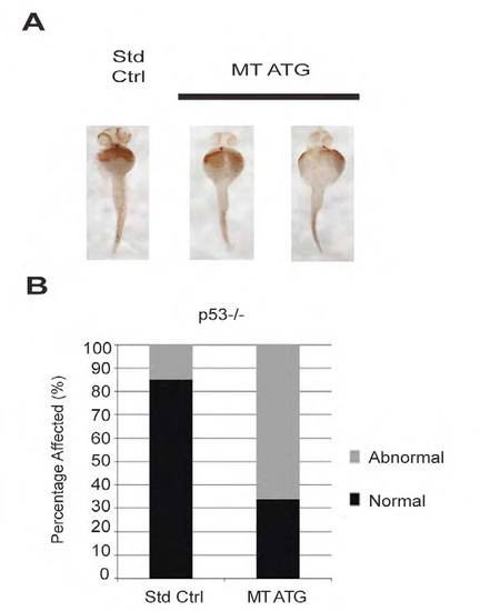
Loss of hemoglobinized red cells in morphant tp53-/- embryos. Morpholinos targeting etv7 were injected into tp53-/- embryos at the single cell stage. Embryos were stained with o-dianisidine at 2 days post fertilization (dpf). B. Quantitation of A. |
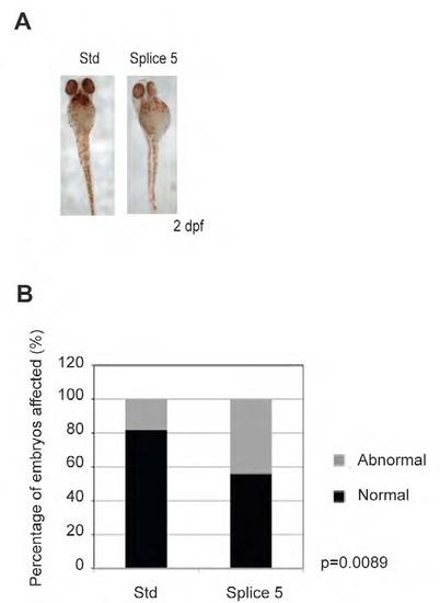
An independent etv7 specific splice inhibiting morpholino causes a significant reduction in hemoglobinized red cells. Wildtype embryos were injected with 8.2 nanograms of standard control morpholino or 8.2 nanograms of a splice site inhibiting morpholino targeting exon 5 of etv7. o-dianisidine staining was performed at 2 days post fertilization (dpf). n= 50 B. Quantitation of A. |

Expression of lss in adult zebrafish tissues. Semi-quantitative RT-PCR of lss mRNA in adult zebrafish tissues. actin is provided as a positive loading control. EXPRESSION / LABELING:
|