
- Title
-
Parallel evolution of chordate cis-regulatory code for development
- Authors
- Doglio, L., Goode, D.K., Pelleri, M.C., Pauls, S., Frabetti, F., Shimeld, S.M., Vavouri, T., and Elgar, G.
- Source
- Full text @ PLoS Genet.

Analysis of Pax6_ciCNE2 and Meis_ciCNE10 in zebrafish embryos. Pax6_ciCNE2 drives GFP expression in both cranial ganglia (A, C) and spinal cord sensory neurons (B, D) at 48 hrs post fertilization. (B, D) High magnification view of GFP expression detectable in neuronal projections extending into the tail fin and along the spinal cord. (E) Meis_ciCNE10 drives GFP expression in the nervous system at 48 hpf. GFP is strongly expressed in the cell body and in the peripheral process of Rohon-Beard neurons innervating the ventral fin fold (F) and the tail fin (G). (H) GFP expression is detected in trigeminal ganglion neurons and in their process innervating the yolk sac (arrow). (I, J) Confocal analysis showing GFP expression in sensory neurons of the trigeminal ganglion innervating the head as well as in central axons innervating the hindbrain (I) and in projections of Rohon-Beard neurons (J). |
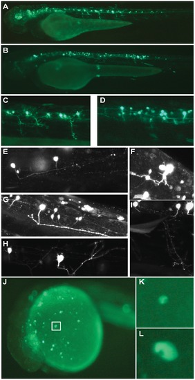
Analysis of Meis_ciCNE1 and Hhex_ciCNE1 in zebrafish embryos. Meis_ciCNE1 drives GFP expression in spinal cord motor neurons (A, C) and interneurons (B, D). Confocal analysis allowed to identify morphological subtypes of interneurons (E–G) and motor neurons (H, I). Intronic Hhex_CNE1 drives GFP expression in a discrete population of blood cell precursors, possibly macrophages (J–L). |
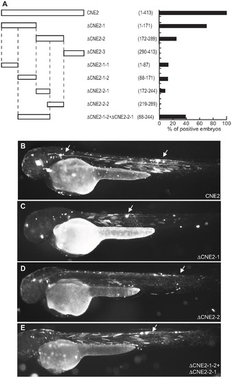
Functional analysis of Pax6_ciCNE2 deletion constructs. (A) Schematic representation and quantification of the constructs injected in the zebrafish embryos to examine their enhancer activity. The numbers in parentheses indicate the length of each construct. (B) View of a 48 hpf embryo injected with the full length CNE2 expressing GFP in sensory neurons (arrow). In the embryos injected with DCNE2-1 (C) or DCNE2-2 (D) GFP is detected in sensory neurons (arrow). The minimal construct DCNE2-1-2+DCNE2-2-1 (E) is able to drive GFP expression in neurons along the spinal cord (arrow). |

Deletion analysis of Meis_ciCNE1 construct. (A) Scheme and quantification of the deletion constructs injected into zebrafish embryos and analysed for GFP expression at 48 hpf. The numbers in parentheses indicate the length of each construct. (B) Embryo injected with the full length CNE1 show GFP expression in the brain and in spinal cord motor- and interneurons. (C) Injection of DCNE1-3 drives GFP expression only in a few neurons in the head. (D) In the embryos injected with DCNE1-2+DCNE1-3 construct few motor- and interneurons are detected along the spinal cord. (E) In embryos injected with the construct DCNE1-4 GFP expression shows a pattern similar to full length, labelling both spinal cord interneurons and motor neurons as well as cells in the brain. |

Deletion analysis of Meis_ciCNE10 construct. (A) Schematic view and quantification of the constructs injected in the embryos and analyzed at 48 hpf. The numbers in parentheses indicate the size of each construct. The green area represents a putative Pbx-Hox site (nt71–79) and the black area a random 10 bp sequence (nt 83–92). (B) View of an embryo injected with the full length CNE10 showing GFP expression in the trigeminal ganglion (arrowhead) and spinal cord sensory neurons (arrow). The two elements DCNE10-2 (C) and DCNE10-2-2 (D, E) retain the same enhancer potential as the full length construct (B). Embryo injected with the 37 bp DCNE10-2oligo1 (F) or with the 24 bp DCNE10-2oligo2 (G, H) show strong GFP expression in sensory neurons (arrow) and trigeminal ganglia (arrowhead). |

Mutational analysis of putative binding site in CNE10. (A) Schematic representation and quantification of mutations introduced in the 24 nt core sequence of CNE10. Mutated triplets are shown in bold. (B) Embryos injected with wt CNE10 show GFP expression in the nervous system at 48 hpf. (C, E) Embryos injected with mut1 and mut 6 show a GFP expression pattern similar to the wt construct, whereas injection of mut5 (D) drives GFP expression only in few neuronal cells. (F) In embryos injected with mut7 GFP expression is mainly detected in trigeminal ganglion neurons. |
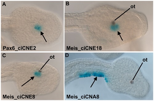
Analysis of ciCNEs in C. intestinalis embryos. (A) Dorsal view of the head of a tailbud embryo electroporated with Pax6_ciCNE2. Reporter expression (arrow, blue stain) is confined to the sensory vesicle. (B) Dorso-lateral view of the head of a tailbud embryo electroporated with Meis_ciCNE10. Reporter expression (arrow) is localised to the sensory vesicle, adjacent to the otolith pigment cell (ot). (C, D) Lateral and dorsal views respectively of tailbud embryos electroporated with Meis_ciCNE1. In (C) reporter expression (arrow) is localised to the sensory vesicle, adjacent to the otolith pigment cell. In (D) reporter expression (arrow) is confined to the lateral tail epidermis of the right hand side. |