
- Title
-
Orthopedia Transcription Factor otpa and otpb Paralogous Genes Function during Dopaminergic and Neuroendocrine Cell Specification in Larval Zebrafish
- Authors
- Fernandes, A.M., Beddows, E., Filippi, A., and Driever, W.
- Source
- Full text @ PLoS One
Expresion of otpa and otpb in wildtype larvae. EXPRESSION / LABELING:
|
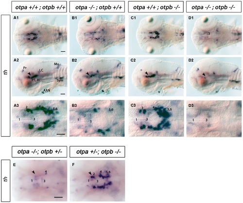
Analysis of DA neurons by expression of th in otpa and otpb single and double mutant larvae. (A–D) Whole-mount in situ hybridization of 3 dpf larvae reveals reduction of th expression in the posterior tuberculum of otpa and total loss of the expression in otpa;otpb double mutants (arrowhead). Other th expressing domains are not affected. (A1–D1, A3–D3) Dorsal views, anterior at left; (A2–D2) lateral views, dorsal up. Scale bar is 50 μm. (E,F) Whole-mount in situ hybridization of 3 dpf larvae reveals reduction of th expression in the posterior tuberculum of otpa mutant, otpb heterozygous larvae (E) (arrowhead). No clear reduction is detected in the posterior tuberculum of otpb mutant, otpa heterozygous larvae (F) (arrowhead). Dorsal view, anterior at left. Scale bar is 50 μm. Abbreviations: AAC, arch associated cluster; DC, diencephalic cluster; H, hypothalamus; LC, locus coeruleus; MO, medulla oblongata; Pr, pretectum; PT, posterior tuberculum. Numbers indicate dopaminergic neurons in the ventral thalamic cluster (1) and posterior tuberculum/hypothalamus (2–6) according to [20]. Scale bar is 50 μm. EXPRESSION / LABELING:
|
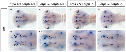
Expression of crh in otpa and otpb single and double mutant larvae. Whole-mount in situ hybridization of 3 dpf larvae reveals loss of crh expression in the preoptic region and posterior tuberculum of otpa;otpb double mutant larvae (arrowhead and asterisk, respectively). Dorsal view, anterior at left. Scale bar is 50 μm. Abbreviations: H, hypothalamus; PO, preoptic region; PT, posterior tuberculum. EXPRESSION / LABELING:
|
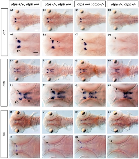
Expression of crh in otpa and otpb single and double mutant larvae. Whole-mount in situ hybridization reveals loss of oxt, avp and trh expression in the preoptic region (arrowhead) of otpa;otpb double mutant larvae at 3 dpf. We detected oxt-expressing cells at ectopic locations within the diencephalon in otpa mutants (B2, asterisk). A reduction of trh-expressing cells in the preoptic region in otpa single mutants is detectable, (J2, arrowhead). Dorsal view, anterior at left. Scale bar is 50 μm. Abbreviations: H, hypothalamus; PO, preoptic region. EXPRESSION / LABELING:
|
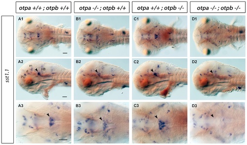
Expression of sst1.1 in otpa and otpb single and double mutant larvae. Whole-mount in situ hybridization reveals reduction of sst1.1 expression (arrowhead) in the rostral hindbrain of otpa mutants and total loss of the expression in otpa;otpb double mutant larvae at 3 dpf. (A1–D1, A3–D3) Dorsal view, anterior at left; (A2–D2) lateral view, dorsal up. Scale bar is 50 μm. Abbreviations: d, diencephalon; HB, hindbrain; m, mesencephalon; PO, preoptic region. EXPRESSION / LABELING:
|
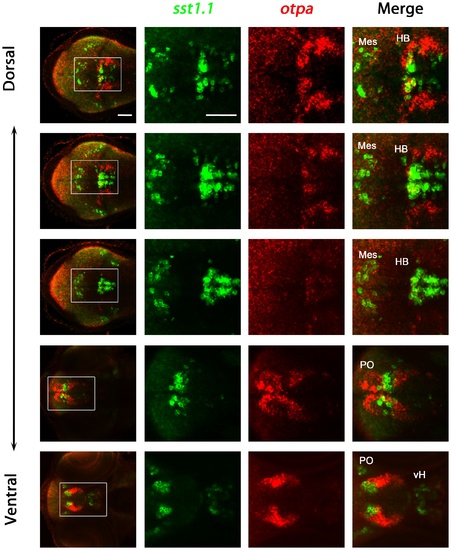
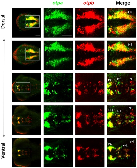
Analysis of coexpression of otpa and sst1.1 in wildtype larvae. Expression of otpa and sst1.1 were detected by double fluorescent whole mount in situ hybridization of wildtype larvae fixed at 3 dpf. From the whole confocal image stack, sub-stacks ranging from dorsal hindbrain image planes to ventral forebrain planes were used to generate a series of dorso-ventral Z-projections. The data reveal that sst1.1 and otpa expression domains overlap in the rostral hindbrain in wildtype larvae at 3 dpf, and some cells appear to coexpress both genes. Dorsal view, anterior at left. Abbreviations: cH, caudal hypothalamus; HB, hindbrain; Mes, mesencephalon; PO, preoptic region; vH, ventral hypothalamus. Scale bar is 50 μm. EXPRESSION / LABELING:
|
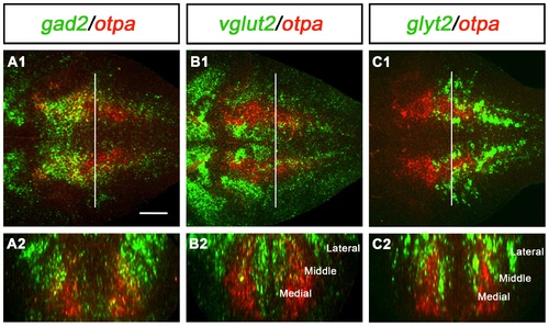
Expression of otpa in relation to gabaergic, glutamatergic and glycinergic markers in the hindbrain of wildtype larvae. Potential coexpression of otpa with gabaergic (gad2, A), glutamatergic (vglut2, B) and glycinergic (glyt2, C) markers was analyzed by double whole mount FISH at 3 dpf. A1, B1, and C1 are single plane dorsal views of the hindbrain, anterior is to the left. A2, B2, and C2 are cross-sections at the level of the hindbrain indicated by the white line in A1, B1, and C1, respectively. The orthogonal view cross sections were obtained from dorsal confocal stacks using the TransformJ Turn plugin of the ImageJ software. Scale bar is 50 μm. EXPRESSION / LABELING:
|
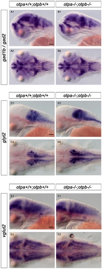
Expression of gad1b/2, glyt2 and vglut2 in wildtype and in otp;otpb double mutant larvae. Whole-mount in situ hybridization reveals no changes in expression of gabaergic (gad1b/gad2), glycinergic (glyt2)and glutamatergic (vglut2) in the hindbrain of otpa;otpb double mutant larvae at 3 dpf. (A1, B1, C1, D1, E1, F1) lateral view, dorsal up; (A2, B2, C2, D2, E2,F2) dorsal view, anterior at left. Scale bar is 50 μm. EXPRESSION / LABELING:
|
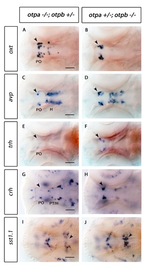
Expression of oxt, avp, trh, crh and sst1.1 in otpa and otpb mutant larvae. Whole-mount in situ hybridization of 3 dpf larvae reveals changes of oxt, avp, trh and crh expression in the preoptic region (arrowhead in A, C, E, G) and reduction of sst1.1 expression (arrowhead in I) in the hindbrain of otpa-/- mutant, otpb+/- heterozygous larvae. In contrast, no obvious change is detected in the preoptic region (arrowheads in B, D, F, H) and hindbrain (J) of otpb-/- mutant, otpa+/- heterozygous larvae. Dorsal view, anterior at left. Scale bar is 50 μm. H, hypothalamus; PO, preoptic region; PT, posterior tuberculum. |
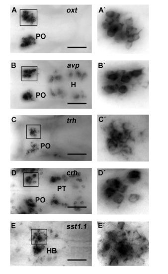
High-resolution imaging of neuroendocrine cells for cell counting. Example of 3 dpf wildtype embryos imaged at single-cell resolution for quantification of cell numbers. (A) oxt, (B), avp, (C) trh, (D) crh expression analysis by WISH. For this figure, from the whole image stack with images at 1 μm spacing, sub-stacks of planes were used to generate a series of dorso-ventral Z-projections containing the region of interest. A higher magnification of regions of interest is shown on the right panel. Dorsal view, anterior at left. Scale bar is 100 μm. H, hypothalamus; HB, Hindbrain; PO, preoptic region; PT, posterior tuberculum. |
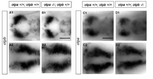
Expression of otp genes is not altered in otp mutant larvae. Whole-mount in situ hybridization of 3 dpf larvae reveals no obvious changes of otpb expression in the preoptic region (A1, B1) and hindbrain (A2, B2) in otpa mutants. Similarly, no obvious changes in otpa expression were detected in the preoptic region (C1, D1) and hindbrain (C2, D2) expression domains in 3 dpf otpb mutants. Scale bar is 100 μm. |