
- Title
-
Treatment of Glucocorticoids Inhibited Early Immune Responses and Impaired Cardiac Repair in Adult Zebrafish
- Authors
- Huang, W.C., Yang, C.C., Chen, I.H., Liu, Y.M., Chang, S.J., and Chuang, Y.J.
- Source
- Full text @ PLoS One
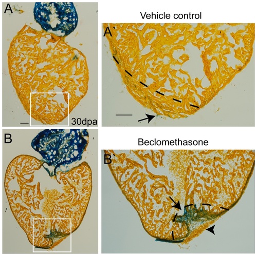
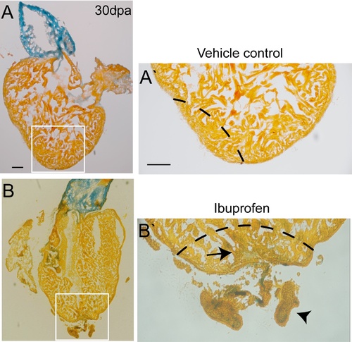
Treatment with beclomethasone caused impaired cardiac repair in zebrafish. Objects were sacrificed at 30 days post ventricular resection; heart sections were stained with aniline blue to distinguish scar tissue from normal tissue. Blue: scar tissue; Orange: normal tissue. (A, A′) In the vehicle control group, the zebrafish regenerated the injured hearts perfectly 1 month after injury. Only a few scar tissue (arrow) was detected in the wound (n = 17). (B, B′) Exposure to beclomethasone (0.25 μM) hindered the cardiac repair process. A large amount of scar tissue (arrow) was found in the wound instead of renewed cardiomyocytes. Meanwhile, obvious blood clots remained, which could be detected (arrow head) (n = 14, scale bar = 100 μm). The dashed lines indicate the approximate amputation plane. |
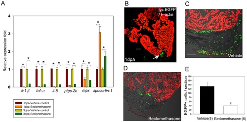
Beclomethasone treatment reduced pro-inflammatory gene expression and phagocyte recruitment after cardiac injury. Whole hearts were harvested at 1 day and 3 days post ventricular resection. RT-qPCR was conducted to quantify the relative fold of pro-inflammatory genes at hearts. (A) Expression of il-1β, tnf-α, il-8, ptgs-2b, and mpx were all significantly reduced in the beclomethasone-treated group for at least 3 days, while lipocortin-1 expression was significantly induced after beclomethasone treatment (n = 3). * indicates p<0.05. (B) Immunostaining of hearts after injury. Green: phagocytes (lyz-EGFP); red: Phalloidin-546. After cardiac injury, numerous phagocytes accumulated in the wound (arrow). (C, E) In the vehicle control group, average of 133±20 phagocytic cells were counted in the fibrin clots (n = 8). (D, E) In the beclomethasone treated animals, 40±5 cells were counted (n = 6). (E) Treatment with beclomethasone significantly impaired phagocyte recruitment in the injured hearts. The data represent the mean±SEM, *indicates p<0.05, scale bar = 100 μm. |
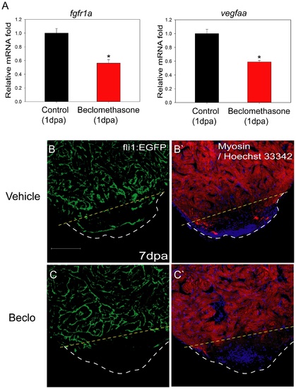
Angiogenesis was hindered in the beclomethasone-treated zebrafish during cardiac repair. We checked pro-angiogenic genes of drug-treated objects at early regeneration and then examined neo-vascularization at 7 dpa. (A) At 1 dpa, mRNA expression of pro-angiogenic genes was reduced after beclomethasone treatment with a 43.7% reduction for fgfr1a and 41% for vegfaa. The data represent the mean±SEM. *indicates p<0.05. (B, B′) At 7 dpa, there were blood vessels appearing in the clotted areas of the control animals. (C, C′) The beclomethasone-treated animals showed little formation of new blood vessels. The yellow dashed lines indicate the approximate amputation plane; the white dashed lines indicate the outline of the apex. Scale bar = 100 μm. |
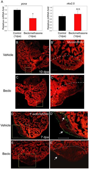
Cell proliferation was hindered in the beclomethasone-treated zebrafish during cardiac repair. Cell proliferation was examined in early phase of regeneration. (A) At 1 dpa, mRNA expression of proliferation marker gene, pcna, was reduced after beclomethasone treatment (40.3% reduction). In contract, the expression of the pre-cardiac gene, nkx2.5, showed no significant difference between the two groups. The data represent the mean±SEM. *indicates p<0.05. N.S. indicates no significant difference. (B, B′) In the control animals, numerous proliferating BrdU+ cells could be detected in the wound (n = 6). The dashed lines indicate the approximate amputation plane. (C, C′) The beclomethasone-treated animals showed little cell proliferation activities (n = 7, scale bar = 50 μm). (D, D′) In control groups, GATA4+ regenerating cells appeared near the injury site at 7 dpa. (arrow, n = 5) (E, E′) Beclomethasone treatment significantly reduced the GATA4+ regenerating cells during cardiac repair (arrow, n = 5, scale bar = 100 μm). The dashed lines indicate the approximate amputation plane. |
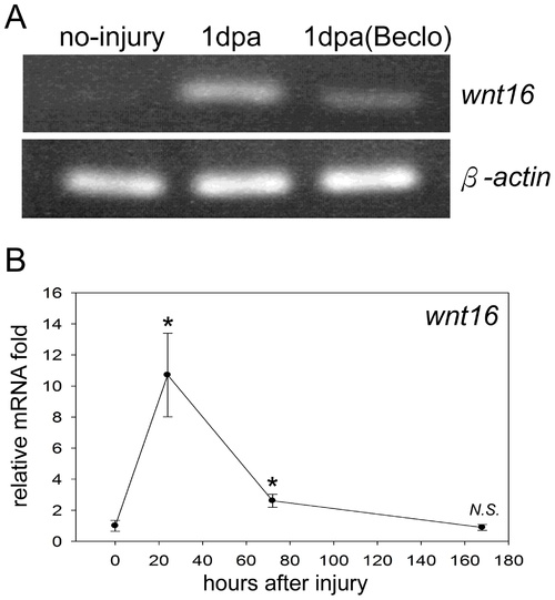
wnt16 was inhibited by beclomethasone in the early phase of zebrafish cardiac repair. RT-PCR assays revealed the normal injury response of wnt16 expression patterns, and confirmed the inhibitory effect of beclomethasone on wnt16 mRNA expression in zebrafish heart. We then checked the mRNA expression of wnt16 before ventricular resection and at 1 dpa, 3 dpa and 7 dpa by RT-qPCR. (A) Zebrafish wnt16 mRNA was elevated at 1 dpa after injury. In contrast, its expression was repressed by beclomethasone treatment at 1 dpa. (B) During zebrafish cardiac repair, wnt16 transcript was highly elevated at 1 dpa. Its level was diminished by 3 dpa, and gradually reduced to the basal level by 7 dpa. (n = 3) The data represent the mean±SEM. *indicates p<0.05. N.S. indicates no significant difference compared to no-injury control. |
Treatment with ibuprofen also caused impaired cardiac repair in zebrafish. Blue: scar tissue; Orange: normal tissue. (A, A′) In the vehicle control group, the zebrafish regenerated the injured hearts perfectly 1 month after injury. (n = 3). (B, B′) Persistent exposure to ibuprofen (0.25 μM) hindered the cardiac repair process. Some scar tissue (arrow) was found in the wound instead of renewed cardiomyocytes; obvious blood clots and cellular debris were left at the wound. (arrow head) (n = 5, scale bar = 100 μm). The dashed lines indicate the approximate amputation plane. |